- Muscle
- Muscle is the tissue of the body which primarily functions as a source of power. There are three types of muscle in the body. Muscle which is responsible for moving extremities and external areas of the body is called "skeletal muscle." Heart muscle is called "cardiac muscle." Muscle that is in the walls of arteries and bowel is called "smooth muscle."
* * *A primary tissue, consisting predominantly of highly specialized contractile cells, which may be classified as skeletal m., cardiac m., or smooth m.; microscopically, the latter is lacking in transverse striations characteristic of the other two types; one of the contractile organs of the body by which movements of the various organs and parts are effected; typical musculus is a mass of musculus fibers (venter or belly), attached at each extremity, by means of a tendon, to a bone or other structure; the more proximal or more fixed attachment is called the origin, the more distal or more movable attachment is the insertion; the narrowing part of the belly that is attached to the tendon of origin is called the caput or head.For gross anatomic description, see musculus. SYN: musculus [TA]. [L. musculus]- muscles of abdomen [TA] muscles forming the wall of the abdomen including rectus abdominis, external and internal oblique muscles, transversus abdominis, and quadratus abdominis. SYN: musculi abdominis.- abductor (m.) [TA] m. that causes movement away from the median plane of body, axis of middle finger, or axis of second toe, or in the case of the thumb, anterior to the plane of the palm. SYN: musculus abductor [TA], abductor.- abductor digiti minimi (m.) of foot [TA] m. of first layer of plantar muscles; origin, lateral and medial processes of calcaneal tuberosity; insertion, lateral side of proximal phalanx of fifth toe; action, abducts and flexes little toe; nerve supply, lateral plantar nerve. SYN: musculus abductor digiti minimi pedis [TA], abductor m. of little toe, musculus abductor digiti quinti (2).- abductor digiti minimi (m.) of hand [TA] superficial hypothenar m. of palm; origin, pisiform bone and pisohamate ligament; insertion, medial side of base of proximal phalanx of the little finger; action, abducts and flexes little finger; nerve supply, deep branch of ulnar. SYN: musculus abductor digiti minimi manus [TA], abductor m. of little finger, musculus abductor digiti quinti (1).- abductor hallucis (m.) [TA] m. of third layer of plantar muscles; origin, medial process of calcaneal tuberosity, flexor retinaculum, and plantar aponeurosis; insertion, medial side of proximal phalanx of great toe; action, abducts great toe; nerve supply, medial plantar. SYN: musculus abductor hallucis [TA], abductor m. of great toe.- abductor pollicis brevis (m.) [TA] superficial thenar m. origin, tubercle of trapezium and flexor retinaculum; insertion, lateral side of proximal phalanx of thumb; action, abducts thumb; nerve supply, median. SYN: musculus abductor pollicis brevis [TA], short abductor m. of thumb.- abductor pollicis longus (m.) [TA] outcropping m. of posterior compartment of forearm; origin, interosseous membrane and posterior surfaces of radius and ulna; insertion, lateral side of base of first metacarpal bone; action, abducts and assists in extending thumb; nerve supply, radial. SYN: musculus abductor pollicis longus [TA], long abductor m. of thumb, musculus extensor ossis metacarpi pollicis.- adductor m. [TA] m. that causes movement toward the median plane of the body, the axis of the third finger or second toe, or, in the case of the thumb, the plane of the palm. SYN: musculus adductor [TA], adductor.- adductor brevis (m.) [TA] m. of medial (adductor) compartment of thigh; origin, superior ramus of pubis; insertion, upper third of medial lip of linea aspera; action, adducts thigh; nerve supply, obturator. SYN: musculus adductor brevis [TA], short adductor m..- adductor hallucis (m.) [TA] m. of third layer of plantar muscles; origin, by two heads, the transverse head from the capsules of the lateral four metatarsophalangeal joints and the oblique head from the lateral cuneiform and bases of the third and fourth metatarsal bones; insertion, lateral side of base of proximal phalanx of great toe; action, adducts great toe; nerve supply, lateral plantar. SYN: musculus adductor hallucis [TA], adductor m. of great toe.- adductor longus (m.) [TA] m. of medial (adductor) compartment of thigh; origin, symphysis and crest of pubis; insertion, middle third of medial lip of linea aspera; action, adducts thigh; nerve supply, obturator. SYN: musculus adductor longus [TA], long adductor m..- adductor magnus (m.) [TA] m. of medial (adductor) compartment of thigh; origin, ischial tuberosity and ischiopubic ramus; insertion, linea aspera and adductor tubercle of femur; action, adducts and extends thigh; nerve supply, obturator and sciatic. SYN: musculus adductor magnus [TA], great adductor m..- adductor minimus (m.) [TA] a small flat m. of the medial (adductor) compartment of thigh constituting the upper portion of the adductor magnus, insertion, the space above linea aspera. SYN: musculus adductor minimus [TA].- adductor pollicis (m.) [TA] intrinsic m. of palm; origin, by two heads, the transverse head from the shaft of the third metacarpal and the oblique head from the front of the base of the second metacarpal, the trapezoid and capitate bones; insertion, medial side of base of proximal phalanx of thumb; action, adducts thumb; nerve supply, ulnar. SYN: musculus adductor pollicis [TA], adductor m. of thumb.- muscles of anal triangle [TA] voluntary muscles of region posterior to perineal body and transverse perineal muscles and anterior to the inferior margins of gluteus maximus muscles; includes levator ani (including puborectalis), and all portions of the external anal sphincter. SYN: musculi regionis analis [TA].- anconeus m. [TA] origin, back of lateral condyle of humerus; insertion, olecranon process and posterior surface of ulna; action, extends forearm and abducts ulna in pronation of wrist; nerve supply, radial. SYN: musculus anconeus [TA], anconeus.- anorectoperineal muscles [TA] smooth m. fibers that pass forward from the longitudinal m. layer of the rectum to the membranous urethra in the male. SYN: musculi anorectoperineales [TA], musculi rectourethrales, rectourethral muscles.- antagonistic muscles two or more muscles that produce opposite movements (function), the contraction of one having the potential, in theory, to “neutralize” that of the other; however, in so doing, they are frequently acting as synergists in fixing the moving part.- anterior cervical intertransversarii (muscles) [TA] deep m. of back; origin, anterior tubercles of cervical transverse processes; insertion, anterior tubercle of next superior transverse process; action, abducts cervical vertebrae; nerve supply, ventral branch of cervical nerves. SYN: musculi intertransversarii anteriores cervicis [TA], anterior cervical intertransverse muscles.- antigravity muscles the muscles that maintain the posture characteristic of a given animal species. In most mammals they are the extensor muscles.- antitragicus (m.) [TA] a band of transverse muscular fibers on the outer surface of the antitragus, arising from the border of the intertragic notch and inserted into the antihelix and tail of the helix. SYN: musculus antitragicus [TA], m. of antitragus.- appendicular m. one of the skeletal muscles of the limbs.- arrector m. of hair [TA] bundles of smooth m. fibers, attached to the deep part of the hair follicles, passing outward alongside the sebaceous glands to the papillary layer of the dermis; they act to pull the hairs erect, causing “goose bumps” or “goose flesh” (cutis anserina) in humans but increasing depth (efficiency) of fur/hair coats of most animals. SYN: musculus arrector pili [TA], arrector pili muscles, erector m. of hair.- articular m. a m. that inserts directly onto the capsule of a joint, acting to retract the capsule in certain movements. SYN: musculus articularis.- articularis cubiti (m.) [TA] the name applied to a small slip of the medial head of the triceps that inserts into the capsule of the elbow joint. SYN: musculus articularis cubiti [TA], articular m. of elbow, subanconeus m..- articularis genus (m.) [TA] deep, distal portion of vastus intermedius m.; origin, lower fourth of anterior surface of shaft of femur; insertion, suprapatellar bursa of knee joint; action, retracts suprapatellar bursa, during extension of knee; nerve supply, femoral. SYN: musculus articularis genus [TA], articular m. of knee, Dupré m., subcrural m., subcruralis, subcrureus, subquadricipital m..- muscles of auditory ossicles [TA] the musculus stapedius and musculus tensor tympani. SYN: musculi ossiculorum auditus [TA], musculi ossiculorum auditoriorum.- auricular muscles [TA] small muscles associated with the auricle, having little function in humans. SYN: musculi auriculares [TA].- auricularis anterior (m.) [TA] origin, epicranial aponeurosis, insertion, cartilage of auricle; action, draws pinna of ear upward and forward; nerve supply, facial. Considered by some to be the anterior part of the temporoparietalis m.. SYN: anterior auricular m., musculus attrahens aurem, musculus attrahens auriculam, musculus auricularis anterior, zygomaticoauricularis.- auricularis posterior (m.) [TA] facial m. of external ear; origin, mastoid process; insertion, posterior portion of root of auricle; action, draws back the pinna; nerve supply, facial. SYN: musculus auricularis posterior [TA], musculus retrahens aurem, musculus retrahens auriculam, posterior auricular (m.).- auricularis superior (m.) [TA] facial m. associated with external ear; origin, galea aponeurotica; insertion, cartilage of auricle; action, draws pinna of ear upward and backward; nerve supply, facial. Considered by some to be the posterior part of the temporoparietal m.. SYN: musculus auricularis superior [TA], attollens aurem, attollens auriculam, musculus attollens aurem, musculus attollens auriculam, superior auricular m..- muscles of back [TA] the muscles of the back in general, including the thoracoappendicular muscles attaching the shoulder girdle to the trunk posteriorly, the posterior serratus muscles, and the erector spinae and transversospinalis muscles. SYN: musculi dorsi [TA], dorsal muscles.- muscles of back proper [TA] muscles of the back innervated by the dorsal primary rami of spinal nerves; includes erector spinae, transversospinalis, interspinales, and anterior and lateral intertransversii muscles; excludes the superficial back muscles which are appendicular and are innervated by ventral rami, and the trapezius, innervated by the spinal accessory nerve. SYN: musculi dorsi proprii [TA], deep muscles of back, true muscles of back.- Bell m. a band of muscular fibers, forming a slight fold in the wall of the bladder, running from the uvula to the opening of the ureter on either side, bounding the trigonum.- biceps brachii (m.) [TA] superficial m. of anterior (flexor) compartment of arm; origin, long head from supraglenoidal tubercle of scapula, short head from coracoid process; insertion, tuberosity of radius; action, flexes elbow and supinates forearm (it is the primary supinator of the forearm); nerve supply, musculocutaneous. SYN: musculus biceps brachii [TA], biceps m. of arm.- biceps femoris (m.) [TA] hamstring m. of posterior compartment of thigh; origin, long head (caput longum) from tuberosity of ischium, short head (caput breve) from lower half of lateral lip of linea aspera; insertion, head of fibula; action, flexes knee and rotates the flexed leg laterally; nerve supply, long head, tibial, short head, fibular. SYN: musculus biceps femoris [TA], biceps m. of thigh, musculus biceps flexor cruris.- bipennate m. SYN: pennate m..- brachialis (m.) [TA] deep m. of anterior (flexor) compartment of arm; origin, lower two-thirds of anterior surface of humerus; insertion, coronoid process of ulna; action, flexes elbow; nerve supply, musculocutaneous, usually with a minor contribution from the radial. SYN: musculus brachialis [TA], brachial m..- brachioradial m. SYN: brachioradialis (m.).- brachioradialis (m.) [TA] m. of posterior (extensor) compartment of forearm; origin, lateral supracondylar ridge of humerus; insertion, anterior aspect of base of styloid process of radius; action, flexes elbow and assists in returning the pronated or supinated limb to the neutral position; nerve supply, (common) radial. SYN: musculus brachioradialis [TA], brachioradial m..- branchiomeric muscles the muscles associated with the branchial arches; they provide a large portion of the musculature for the face and neck; the myoblasts for these muscles originate from paroxial mesoderm, while the neural crest provides their connective tissue.- bronchoesophageal m. SYN: bronchoesophageus (m.).- bronchoesophageus (m.) [TA] muscular fascicles, arising from the wall of the left bronchus, which reinforce the musculature of the esophagus. SYN: musculus bronchoesophageus [TA], bronchoesophageal m..- buccinator (m.) [TA] facial m. of cheek; origin, posterior portion of alveolar portion of maxilla and mandible and pterygomandibular raphe; insertion, angle of mouth; also become interspersed with more horizontal portions of the orbicularis oris; action, flattens cheek, retracts angle of mouth; plays an important role in mastication, working with tongue and orbicularis oris m. to keep food between teeth; when it is paralyzed, as in Bell palsy, food accumulates in the oral vestibule; nerve supply, facial. SYN: musculus buccinator [TA], cheek m..- bulbocavernosus m. SYN: bulbospongiosus (m.).- bulbospongiosus (m.) [TA] perineal m.; in the male : origin, the perineal membrane fascia on the dorsum of the bulb of the penis; insertion, central tendon of the perineum and the median raphe on the free surface of the bulb; action, voluntarily constricts bulbous urethra when attempting to expel last drops following urination, or spasmodically with, and following, ejaculation to expel semen. In the female : origin, the dorsum of the clitoris, the corpus cavernosum, and the perineal membrane; insertion, central tendon of the perineum; action, acts as a weak sphincter of the vagina; when developed, is a part of “cross-member musculature” of pelvic floor that resists prolapse of pelvic viscera; surrounds and compresses greater vestibular gland, especially during erection of bulb of vestibule, expressing secretion. Nerve supply, pudendal (deep perineal branch). SYN: musculus bulbospongiosus [TA], bulbocavernosus m., musculus bulbocavernosus, musculus ejaculator seminis, musculus sphincter vaginae, sphincter vaginae.- cardiac m. the involuntary m. comprising the myocardium and walls of the pulmonary veins and superior vena cava, consisting of anastomosing transversely striated m. fibers formed of cells united at intercalated disks; the one or two nuclei of each cell are centrally located and the longitudinally arranged myofibrils have considerable sarcoplasm around them; connective tissue is limited to reticular and fine collagenous fibers; contraction is rhythmic and intrinsically stimulated. SYN: m. of heart.- Casser perforated m. SYN: coracobrachialis m..- ceratocricoid (m.) [TA] an inconstant fasciculus from the posterior cricoarytenoid m. inserted into the inferior horn of the thyroid cartilage. SYN: musculus ceratocricoideus [TA], Merkel m..- ceratoglossus (m.) [TA] main, posterior part of hyoglossus m. (vs. chondroglossus) arising from the greater horn of the hyoid bone. SYN: musculus ceratoglossus [TA].- cervical interspinal m. SYN: interspinales cervicis (muscles).- cervical interspinales muscles SYN: interspinales cervicis (muscles).- chondroglossus m. [TA] lesser part of hyoglossus, arising as fibers from lesser horn of hyoid bone, which are separated from the main part of the hyoglossus (the ceratoglossus) by slips of genioglossus m.. SYN: musculus chondroglossus.- ciliary m. [TA] the intrinsic smooth m. of the ciliary body of eyeball; it consists of circular fibers [TA] (fibrae circulares [TA]), radial fibers [TA] (fibrae radialis [TA]), meridional fibers [TA] (fibrae meridoneales [TA]), and longitudinal fibers [TA] (fibrae longitudinales [TA]); action, in contracting, its diameter is reduced (like a sphincter's), reducing tensile (stretching) forces on lens, allowing the lens to thicken for near vision (accommodation). SYN: musculus ciliaris [TA], Bowman m., ciliary ligament.- coccygeus m. [TA] striated pelvic m. associated with the deep (pelvic) aspect of the sacrospinous ligament, forming part of the pelvic diaphragm; origin, spine of ischium and sacrospinous ligament; insertion, sides of lower part of sacrum and upper part of coccyx; action, with the sacrospinous ligament assists in support of pelvic floor, theoretically increasingly so when intra-abdominal pressures increase; nerve supply, third and fourth sacral. SYN: musculus coccygeus [TA], coccygeal m., ischiococcygeus, musculus ischiococcygeus.- muscles of coccyx the muscles of the coccyx considered as a group, including the musculus coccygeus and the inconstant ventral and dorsal sacrococcygeal muscles. SYN: musculi coccygei.- Coiter m. SYN: corrugator supercilii (m.).- compressor urethra (m.) [TA] part of the external urethral sphincter complex of m. consisting of a slender muscular band spanning between the more posterior aspects of the ischiopubic rami but looping anterior to the urethra so that its contraction pulls posteriorly on the urethra,, compressing its anterior wall against the posterior wall, closing the urethra like a gentle kink in a hose; Terminologia Anatomica lists this m. only for the female, but a similar structure has been described for the male. SYN: musculus compressor urethrae [TA].- coracobrachial m. SYN: coracobrachialis m..- coracobrachialis m. [TA] m. of anterior (flexor) compartment of arm; origin, coracoid process of scapula; insertion, middle of medial border of humerus; action, adducts and flexes the arm; acts as a shunt m. in resisting downward dislocation of shoulder joint; nerve supply, musculocutaneous. SYN: musculus coracobrachialis [TA], Casser perforated m., coracobrachial m..- corrugator cutis m. of anus m. of anal triangle with m. fibers radiating from the superficial portion of the external sphincter to the deep aspect of the perianal skin, said to cause puckering of that skin, which contributes to the air/water-tight “seal” of the anal canal. SYN: musculus corrugator cutis ani.- corrugator supercilii (m.) [TA] facial m. of forehead; origin, from orbital portion of musculus orbicularis oculi and nasal prominence; insertion, skin of eyebrow; action, draws medial end of eyebrow downward and wrinkles forehead vertically, conveying expression of deep thought, worry, or concern; nerve supply, facial. SYN: musculus corrugator supercilii [TA], Coiter m., corrugator m., wrinkler m. of eyebrow.- Crampton m. SYN: Brücke m..- cremaster m. [TA] origin, continuation of fibers from inferiormost internal oblique m. and slips arising from the inguinal ligament; insertion, becomes interspersed within cremasteric fascia of spermatic cord and intermediate covering of testis; in the female, the round ligament of the uterus; action, elevate testicle; nerve supply, genital branch of genitofemoral. SYN: musculus cremaster [TA], Riolan m. (2).- cricothyroid m. [TA] intrinsic laryngeal m.; origin, anterior surface of arch of cricoid; insertion, the anterior or straight part passes upward to ala of thyroid; the posterior or oblique part passes more outward to inferior horn of thyroid; action, acts at cricothyroid joint, pulling anterior aspects of thyroid and cricoid cartilages together, rotating the superior portion of the cricoid lamina and arytenoid cartilages posteriorly, causing vocal folds to tense, increasing the pitch of voice tone; the antagonist of this movement is the thyroarytenoid m.; nerve supply, external laryngeal branch of superior laryngeal nerve (from vagus). SYN: musculus cricothyroideus [TA].- cruciate m. a general type of m. in which the muscles or bundles of m. fibers cross in an X-shaped configuration; e.g., the oblique arytenoid muscles. SYN: musculus cruciatus.- cutaneous m. [TA] a m. that lies in the subcutaneous tissue and attaches to the skin; it may or may not have a bony attachment. The muscles of expression are the chief examples of cutaneous muscles in the human. SYN: musculus cutaneus [TA].- dartos m. smooth m. fibers interspersed within the dartos fascia (superficial fascia of scrotum), causing contraction of the scrotum, as when experiencing a cool environmental temperature. SEE ALSO: dartos fascia.- deep transverse perineal m. [TA] origin, ramus of ischium; insertion, with its fellow in the perineal body; action, with superficial transverse perineal m. in forming the transverse element of cross-member (the sagittal element being formed by bulbospongiosus and external anal sphincter muscles) that provides support of the perineum and the pelvic diaphragm above it during increased abdominopelvic pressure; in males, it adds support to the bulb of the penis; nerve supply, pudendal (dorsal nerve of penis/clitoris). SYN: musculus transversus perinei profundus [TA], deep transverse m. of perineum.- deltoid (m.) [TA] intrinsic (scapulohumeral) m. of shoulder joint; origin, lateral third of anterior border of clavicle, lateral and posterior border of acromion process, lower border of spine of scapula; insertion, lateral side of shaft of humerus (deltoid tuberosity) a little above its middle; action, its anterior, middle, and posterior portions act independently to produce abduction, flexion, extension, and rotation of the humerus at the shoulder joint; nerve supply, axillary (fifth and sixth cervical spinal cord segments via the brachial plexus). SYN: musculus deltoideus [TA].- depressor anguli oris (m.) [TA] facial m. of mouth; origin, anterolateral base of mandible anteriorly; insertion, blends with other muscles in lower lip near angle of mouth; action, pulls down corners of mouth; nerve supply, facial. SYN: musculus depressor anguli oris [TA], musculus triangularis (2) [TA], triangular m. (2) [TA], musculus triangularis labii inferioris.- depressor labii inferioris (m.) [TA] facial m. of mouth; origin, anterior portion of base of mandible; insertion, interdigitates with fibers of orbicularis oris musculus to reach skin of lower lip; action, depresses lower lip; nerve supply, facial. SYN: musculus depressor labii inferioris [TA], depressor m. of lower lip, musculus quadratus labii inferioris, musculus quadratus menti.- depressor septi nasi (m.) [TA] facial m. of nose; a vertical fasciculus from the maxilla superior to the central incisor passing upward along the median line of the upper lip to insert into the mobile part of the nasal septum; action, works with alar (dilator) part of the nasalis m. to widen the nares during deep inspiration; depresses septum; nerve supply, buccal branch of facial. SYN: musculus depressor septi [TA], depressor (m.) of septum.- depressor supercilii (m.) [TA] facial m. arising from nasal part of frontal bone, medial to the corrugator supercili and inserting into the skin underlying the middle of the eyebrow; action, depresses eyebrow; nerve supply, facial. SYN: musculus depressor supercilii [TA], depressor m. of eyebrow.- detrusor (m.) [TA] the muscular coat of the urinary bladder, which, along with gravity and increased intraabdominal pressure, facilitates emptying of bladder during urination by its contraction. SYN: musculus detrusor urinae [TA].- digastric (m.) [TA] 1. one of the suprahyoid group of muscles consisting of two bellies united by a central tendon that passes through a fascial loop connected to the body of the hyoid bone; origin, by posterior belly from the digastric groove medial to the mastoid process; insertion, by anterior belly into lower border of mandible near midline; action, elevates the hyoid when mandible is fixed; depresses the mandible when hyoid is fixed; nerve supply, posterior belly from facial, anterior belly by nerve to the mylohyoid from the mandibular division of trigeminal; SYN: musculus biventer [TA], musculus digastricus [TA], two-bellied m. [TA], biventer mandibulae, musculus biventer mandibulae. 2. a m. with two fleshy bellies separated by a fibrous insertion;- dilator m. [TA] a m. that opens an orifice or dilates the lumen of an organ; it is the dilating or opening component of a pylorus (the other component is the sphincter m.). SYN: musculus dilatator [TA], musculus dilator.- dilator (m.) of ileocecal sphincter the longitudinal muscular fibers that open the ileal orifice at the level of the cecocolic junction. SYN: musculus dilator pylori ilealis.- dilator pupillae m. [TA] intrinsic “m.” of eyeball; radially disposed layer of muscular processes of the myoepithelial cells that form the epithelium of the posterior surface of the iris, which extends from the pupillary to the ciliary margin; sympathetic stimulation causes contraction, which slowly dilates the pupil to allow more light to reach the retina. SYN: musculus dilator pupillae [TA], dilator iridis, dilator of pupil, musculus dilator iridis.- dilator (m.) of pylorus the longitudinal muscular fibers that open the gastroduodenal junction. SYN: musculus dilator pylori gastroduodenalis.- dorsal interossei (interosseous muscles) of foot [TA] four intrinsic muscles of the fourth layer of plantar muscles; origin, from sides of adjacent metatarsal bones; insertion, first into medial, second into lateral side of proximal phalanx of second toe, third and fourth into lateral side of proximal phalanx of third and fourth toes; action, abduct toes 2–4 from an axis through the second toe; nerve supply, lateral plantar. SYN: musculi interossei dorsalis pedis [TA].- dorsal interossei (interosseous muscles) of hand [TA] four intrinsic muscles of the hand; origin, sides of adjacent metacarpal bones; insertion, proximal phalanges and extensor expansion, first on radial side of index, second on radial side of middle finger, third on ulnar side of middle finger, fourth on ulnar side of ring finger; action, abduct fingers 2–4 from the axis of the middle finger, nerve supply, ulnar. SYN: musculi interossei dorsalis manus [TA].- dorsal sacrococcygeus m. an inconstant and poorly developed m. on the dorsal surfaces of the sacrum and coccyx, the remains of a portion of the tail musculature of lower animals. SYN: dorsal sacrococcygeal m., musculus extensor coccygis, musculus sacrococcygeus dorsalis, musculus sacrococcygeus posterior.- elevator m. of rib SYN: levatores costarum (muscles).- epicranius (m.) [TA] compound facial (scalp) m. composed of the epicranial aponeurosis and the muscles inserting into it, i.e., the occipitofrontalis musculus and temporoparietalis musculus. SYN: musculus epicranius [TA], epicranial m., scalp m..- erector spinae (muscles) [TA] proper muscles of back; origin, from sacrum, ilium, and spines of lumbar vertebrae; it divides into three columns, iliocostalis musculus, longissimus musculus, and spinalis musculus, which insert into ribs and vertebrae with additional m. slips joining the columns at successively higher levels; action, extends and laterally flexes vertebral column; nerve supply, dorsal primary rami of spinal nerves. SYN: musculus erector spinae [TA], erector m. of spine, musculus sacrospinalis.- extensor m. [TA] m. producing extension, i.e., a movement that produces straightening, or an increase in the angle of a joint. SYN: musculus extensor [TA].- extensor carpi radialis brevis (m.) [TA] m. of posterior compartment of forearm; origin, lateral epicondyle of humerus; insertion, base of third metacarpal bone; action, extends and abducts hand at wrist joint; nerve supply, deep radial. SYN: musculus extensor carpi radialis brevis [TA], short radial extensor m. of wrist.- extensor carpi radialis longus (m.) [TA] m. of posterior (extensor) compartment of forearm; origin, lateral supracondylar ridge of humerus; insertion, posterior aspect of base of second metacarpal bone; action, extends and abducts hand at wrist joint; nerve supply, radial. SYN: musculus extensor carpi radialis longus [TA], long radial extensor m. of wrist.- extensor carpi ulnaris (m.) [TA] m. of posterior (extensor) compartment of forearm; origin, lateral epicondyle of humerus (humeral head) and oblique line and posterior border of ulna (ulnar head); insertion, base of fifth metacarpal bone; action, extends and adducts hand at wrist joint; nerve supply, radial (posterior interosseous). SYN: musculus extensor carpi ulnaris [TA], ulnar extensor (m.) of wrist.- extensor digiti minimi (m.) [TA] m. of posterior (extensor) compartment of forearm; origin, lateral epicondyle of humerus; insertion, dorsum of proximal, middle, and distal phalanges of little finger; action, extends little finger; nerve supply, radial (posterior interosseous). SYN: musculus extensor digiti minimi [TA], extensor (m.) of little finger, musculus extensor digiti quinti proprius, musculus extensor minimi digiti.- extensor digitorum m. [TA] m. of posterior (extensor) compartment of forearm; origin, lateral epicondyle of humerus; insertion, by four tendons into the base of the proximal and middle and base of the distal phalanges; action, extends fingers, especially at metacarpophalangeal joint; nerve supply, radial (posterior interosseous). SYN: musculus extensor digitorum [TA], extensor (m.) of fingers, musculus extensor digitorum communis.- extensor digitorum brevis (m.) [TA] intrinsic m. of dorsum of foot; origin, dorsal surface of calcaneus; insertion, by four tendons fusing with those of the extensor digitorum longus, and by a slip attached independently to the base of the proximal phalanx of the great toe; action, extends four lateral toes (II–V); nerve supply, deep fibular. SYN: musculus extensor digitorum brevis [TA], musculus extensor brevis digitorum, short extensor (m.) of toes.- extensor digitorum brevis (m.) of hand a short extensor m. of the fingers of rare occurrence, and comparable to the short extensor of the toes. SYN: musculus extensor digitorum brevis manus, Pozzi m..- extensor digitorum longus (m.) [TA] m. of anterior (extensor/dorsiflexor) compartment of leg; origin, lateral condyle of tibia, upper two-thirds of anterior margin of fibula; insertion, by four tendons to the dorsal surfaces of the bases of the proximal, middle, and distal phalanges of the second to fifth toes; action, extends the four lateral toes; nerve supply, deep fibular. SYN: musculus extensor digitorum longus [TA], long extensor (m.) of toes, musculus extensor longus digitorum.- extensor hallucis brevis (m.) [TA] intrinsic m. of dorsum of foot considered by some anatomists as the medial belly of extensor digitorum brevis m.; origin, dorsal calcaneus; insertion, via a stout aspect of the base of the proximal phalanx of the great toe; action, extends great toe; nerve supply, deep fibular. of which is inserted into the base of the proximal phalanx of the great toe. SYN: musculus extensor hallucis brevis [TA], short extensor (m.) of great toe.- extensor hallucis longus (m.) [TA] m. of anterior (extensor/dorsiflexor) compartment of leg; origin, anterior surface of fibula and interosseous membrane; insertion, dorsal aspect of base of distal phalanx of great toe; action, extends the great toe; nerve supply, deep fibular. SYN: musculus extensor hallucis longus [TA], long extensor (m.) of great toe.- extensor indicis (m.) [TA] m. of posterior (extensor) compartment of forearm; origin, dorsal surface of distal ulna and adjacent interosseous membrane; insertion, extensor expansion of index finger; action, independently extends the index finger and assists in extending hand at wrist joint; nerve supply, radial (posterior interosseous). SYN: musculus extensor indicis [TA], index extensor (m.), musculus extensor indicis proprius.- extensor pollicis brevis (m.) [TA] m. of posterior (extensor) compartment of forearm; origin, dorsal surface of distal radius and adjacent interosseous membrane; insertion, posterior aspect of base of proximal phalanx of thumb; action, extends and abducts the thumb at metacarpophalangeal joint; nerve supply, radial joint (posterior interosseous). SYN: musculus extensor pollicis brevis [TA], musculus extensor brevis pollicis, short extensor (m.) of thumb.- extensor pollicis longus (m.) [TA] m. of posterior (extensor) compartment of forearm; origin, posterior surface of middle of shaft of ulna; insertion, dorsal aspect of base of distal phalanx of thumb; action, extends distal phalanx of thumb; nerve supply, radial (posterior interosseous). SYN: musculus extensor pollicis longus [TA], long extensor (m.) of thumb, musculus extensor longus pollicis.- external intercostal (m.) [TA] flat m. of thorax arising from lower border of one rib and pass obliquely downward and forward to be inserted into the upper border of rib below; action, contract during inspiration to elevate ribs; also to maintain tension in the intercostal spaces to resist inward movement during inspiration; nerve supply, intercostal. SYN: musculus intercostales externi [TA].- external oblique (m.) [TA] flat m. of abdomen; origin, external surfaces of fifth to twelfth ribs; insertion, anterior half of lateral lip of iliac crest and inguinal ligament inferiorly, and continuing medially as part of the anterior layer of the rectus sheath; action, supports and compresses abdominal viscera; flexes and rotates trunk; nerve supply, thoracoabdominal nerves. SYN: musculus obliquus externus abdominis [TA], abdominal external oblique (m.).- extraocular muscles [TA] the muscles within the orbit but outside of eyeball, including the four rectus muscles (superior, inferior, medial and lateral); two oblique muscles (superior and inferior), and the levator of the superior eyelid (levator palpebrae superioris). SYN: musculi externi bulbi oculi [TA], extrinsic muscles of eyeball, muscles of eyeball, musculi bulbi, ocular muscles.- extrinsic muscles muscles arising outside of, but which act upon, the structure under consideration. For example, the muscles operating the hand but having fleshy bellies located in the forearm.- muscles of eyeball SYN: extraocular muscles.- facial muscles [TA] the numerous muscles supplied by the facial nerve that are attached to and move the skin of the face. Terminologia Anatomica includes the buccinator m. in this group because of its innervation and embryonic origin, even though it functions primarily in mastication. SYN: mimetic muscles, muscles of facial expression, musculi faciei.- fibularis brevis (m.) [TA] origin, lower two-thirds of lateral surface of fibula; insertion, base of fifth metatarsal bone; action, everts foot; nerve supply, superficial peroneal. SYN: musculus fibularis brevis [TA], musculus peroneus brevis, peroneus brevis (m.), short fibular m., short peroneal m..- fibularis longus (m.) [TA] origin, upper two-thirds of outer surface of fibula and lateral condyle of tibia; insertion, by tendon passing behind lateral malleolus and across sole of foot to medial cuneiform and base of first metatarsal; action, plantar flexes and everts foot; nerve supply, superficial peroneal. SYN: musculus fibularis longus [TA], musculus peroneus longus, peroneus longus (m.), long fibular m., long peroneal m..- fibularis tertius (m.) [TA] origin, in common with musculus extensor digitorum longus; insertion, dorsum of base of fifth metatarsal bone; nerve supply, deep branch of peroneal; action, assists in dorsiflexion and eversion of foot. SYN: musculus fibularis tertius [TA], musculus peroneus tertius, peroneus tertius (m.), third peroneal m..- fixator m. a m. that acts as a stabilizer of one part of the body during movement of another part.- flat m. [TA] broad, relatively thin, sheetlike m., e.g., muscles of the anterolateral abdominal wall (external and internal oblique, transversus abdominis). SYN: musculus plana [TA].- flexor m. [TA] m. producing flexion, i.e., a movement that bends or decreases the angle of joints. SYN: musculus flexor [TA].- flexor carpi radialis (m.) [TA] m. of the anterior (flexor) compartment of forearm; origin, common flexor origin of medial condyle of humerus; insertion, anterior surface of the base of the second and most often sending a slip to that of the third metacarpal bone; action, flexes and abducts wrist; nerve supply, median; its tendon travels in its own canal roofed by a layer of the transverse carpal ligament. SYN: musculus flexor carpi radialis [TA], radial flexor (m.) of wrist.- flexor carpi ulnaris (m.) [TA] m. of anterior (flexor) compartment of forearm; origin, via a humeral head from the medial condyle of humerus and an ulnar head from the olecranon and upper three-fifths of posterior border of ulna; insertion, pisiform bone, but is continued to the fifth metacarpal bone via the pisometacarpal ligament; action, flexes and adducts the hand at the wrist joint ulnarward; nerve supply, ulnar. SYN: musculus flexor carpi ulnaris [TA], ulnar flexor (m.) of wrist.- flexor digiti minimi brevis (m.) of foot [TA] m. of third layer of plantar muscles; origin, base of metatarsal bone of the little toe and sheath of fibularis longus m.; insertion, lateral surface of base of proximal phalanx of little toe; action, flexes the little toe at the metacarpophalangeal joint; nerve supply, lateral plantar. SYN: musculus flexor digiti minimi brevis pedis [TA], short flexor (m.) of little toe.- flexor digiti minimi brevis (m.) of hand [TA] hypothenar m. of palm; origin, hook of hamate bone; insertion, medial side of proximal phalanx of little finger; action, flexes little finger at the metacarpophalangeal joint; nerve supply, deep branch of ulnar. SYN: musculus flexor digiti minimi brevis manus [TA], short flexor (m.) of little finger.- flexor digitorum brevis (m.) [TA] m. of first layer of lantar muscles; origin, medial tubercle of calcaneus and plantar aponeurosis; insertion, middle phalanges of four lateral toes by tendons perforated by those of the flexor digitorum longus; action, flexes lateral four toes; nerve supply, medial plantar. SYN: musculus flexor digitorum brevis [TA], musculus flexor brevis digitorum, short flexor (m.) of toes.- flexor digitorum longus (m.) [TA] m. of deep posterior or flexor (plantar flexor) comparment of leg; origin, middle third of posterior surface of tibia; insertion, by four tendons, perforating those of the flexor brevis, into bases of distal phalanges of four lateral toes; action, flexes second to fifth toes; nerve supply, tibial nerve. SYN: musculus flexor digitorum longus [TA], long flexor (m.) of toes, musculus flexor longus digitorum.- flexor digitorum profundus (m.) [TA] m. of deep layer of anterior (flexor) compartment of forearm; origin, anterior surface of proximal third of ulna; insertion, by four tendons, piercing those of the flexor digitorum superficialis, into anterior aspect of the base of distal phalanx of each finger; action, flexes distal interphalangeal joint of fingers; nerve supply, ulnar and median (anterior interosseous m.). SYN: musculus flexor digitorum profundus [TA], deep flexor (m.) of fingers, musculus flexor profundus.- flexor digitorum superficialis (m.) [TA] intermediate m. of anterior (flexor) compartment of forearm; origin, via a humeroulnar head from the medial epicondyle of the humerus, the medial border of the coronoid process, and a tendinous arch between these points, and a radial head from the oblique line and middle third of the lateral border of the radius; insertion, by four split tendons, passing to either side of the flexor digitorum profundus tendons, into sides of middle phalanx of each finger; action, flexes proximal interphalangeal joint of the fingers; nerve supply, median. SYN: musculus flexor digitorum superficialis [TA], musculus flexor digitorum sublimis, musculus flexor sublimis, superficial flexor (m.) of fingers.- flexor hallucis brevis (m.) [TA] thenar m. of palm; origin, medial surface of cuboid and middle and lateral cuneiform bones; insertion, by two tendons, embracing that of the flexor longus hallucis, into the sides of the base of the proximal phalanx of the great toe; action, flexes great toe; nerve supply, medial and lateral plantar. SYN: musculus flexor hallucis brevis [TA], musculus flexor brevis hallucis, short flexor (m.) of great toe.- flexor hallucis longus (m.) [TA] m. of deep posterior (plantar flexor) compartment of leg; origin, lower two-thirds of posterior surface of fibula; insertion, base of distal phalanx of great toe; action, flexes great toe; nerve supply, medial plantar. SYN: musculus flexor hallucis longus [TA], long flexor (m.) of great toe, musculus flexor longus hallucis.- flexor pollicis brevis (m.) [TA] thenar m. of palm; origin, superficial portion from flexor retinaculum of wrist, deep portion from ulnar side of first metacarpal bone; insertion, palmar aspect of base of proximal phalanx of thumb; action, flexes proximal phalanx of thumb; nerve supply, median (superficial head) and deep branch of ulnar (deep head). Some authors consider the deep head to be the first in a series of four palmar interossei muscles of the hand. SYN: musculus flexor pollicis brevis [TA], short flexor (m.) of thumb.- flexor pollicis longus (m.) [TA] m. of deep layer of anterior (flexor) compartment of forearm; origin, anterior surface of middle third of radius; insertion, palmar aspect of distal phalanx of thumb; action, flexes thumb at interphalangeal joint; nerve supply, median (anterior interosseous). SYN: musculus flexor pollicis longus [TA], long flexor m. of thumb, musculus flexor longus pollicis.- four-headed m. [TA] having four heads; denoting a m. of the thigh, quadriceps femoris m., and—uncommonly—one of the calf, quadriceps surae m., or the combined gastrocnemius (with two heads), soleus, and plantaris, more commonly called triceps surae m., the plantaris being counted as a separate m.. SYN: quadriceps.- fusiform m. [TA] one that has a fleshy belly, tapering at either extremity. SYN: musculus fusiformis [TA], spindle-shaped m..- Gantzer m. an accessory m. extending from the superficial flexor of the digits to the deep flexor of the digits.- gastrocnemius (m.) [TA] superficial m. of posterior (plantar flexor) compartment of leg; origin, by two heads (lateral and medial) from the lateral and medial condyles of the femur; insertion, with soleus by tendo calcaneus into lower half of posterior surface of calcaneus; action, plantar flexion of foot; nerve supply, tibial. SYN: musculus gastrocnemius [TA], gastrocnemius.- genioglossal m. SYN: genioglossus (m.).- genioglossus (m.) [TA] one of the paired lingual muscles; origin, mental spine of the mandible; insertion, lingual fascia beneath the mucous membrane and epiglottis; action, depresses and protrudes the tongue; nerve supply, hypoglossal. SYN: musculus genioglossus [TA], genioglossal m., genioglossus, musculus geniohyoglossus.- geniohyoid (m.) [TA] one of the suprahyoid muscles of the neck; origin, mental spine of mandible; insertion, body of hyoid bone; action, draws hyoid forward, or depresses jaw when hyoid is fixed; nerve supply, fibers from ventral primary rami of first and second cervical spinal nerves conveyed by the hypoglossal nerve. SYN: musculus geniohyoideus [TA], geniohyoid, geniohyoideus.- gluteus maximus (m.) [TA] superficial m. of buttock; origin, ilium behind posterior gluteal line, posterior surface of sacrum and coccyx, and sacrotuberous ligament; insertion, iliotibial band of fascia lata (superficial three-quarters) and gluteal ridge (deep inferior one-quarter) of femur; action, extends thigh, especially from the flexed position, as in climbing stairs or rising from a sitting position; nerve supply, inferior gluteal. SYN: musculus gluteus maximus [TA].- gluteus medius (m.) [TA] intermediate m. of buttock; origin, ilium between anterior and posterior gluteal lines; insertion, lateral surface of greater trochanter; action, abducts and medially rotates thigh; nerve supply, superior gluteal. SYN: musculus gluteus medius [TA], mesogluteus.- gluteus minimus (m.) [TA] deep m. of buttock; origin, ilium between anterior and inferior gluteal lines; insertion, greater trochanter of femur; action, abducts and medially rotates thigh; nerve supply, superior gluteal. SYN: musculus gluteus minimus [TA].- gracilis (m.) [TA] m. of medial compartment of thigh; origin, ramus of pubis near symphysis; insertion, shaft of tibia below medial tuberosity (see pes anserinus); action, adducts thigh, flexes knee, rotates leg medially; nerve supply, obturator. SYN: musculus gracilis [TA], gracilis (2).- hamstring muscles the muscles at the back of the thigh, comprising the long head of biceps, the semitendinosus, and the semimembranosus muscles; hamstring muscles ariose from ischial tuberosity, act across both hip and knee joints, and are innervated by the tibial nerve.- muscles of head [TA] the muscles of expression, of mastication, and the suboccipital muscles in general. SYN: musculi capitis [TA].- helicis major (m.) [TA] auricular m. occurring as a narrow band of muscular fibers on the anterior border of the helix of the auricle, arising from the spine and inserted at the point where the helix becomes transverse. SYN: musculus helicis major [TA], large m. of helix.- helicis minor (m.) [TA] auricular m. occurring as a band of oblique fibers covering the crus of the helix of the auricle. SYN: musculus helicis minor [TA], smaller m. of helix.- hyoglossus (m.) [TA] m. of tongue; origin, body and greater horn of hyoid bone; insertion, side of the tongue; action, retracts and pulls down side of tongue; nerve supply, motor by hyoglossal, sensory by lingual. SYN: musculus hyoglossus [TA], hyoglossal m., hyoglossus.- iliacus (m.) [TA] origin, iliac fossa; insertion, via a common tendon with psoas major into anterior surface of lesser trochanter of the femur, and capsule of hip joint; action, flexes thigh and rotates it medially; nerve supply, lumbar plexus. SYN: musculus iliacus [TA], iliac m..- iliacus minor (m.) the fibers of the iliacus arising from the anterior inferior iliac spine and inserted into the iliofemoral ligament, sometimes distinctly separate from the rest of the m.. SYN: musculus iliacus minor, musculus iliocapsularis.- iliococcygeal m. SYN: iliococcygeus (m.).- iliococcygeus (m.) [TA] the posterior part of the levator ani; origin, tendinous arch of the levator ani m. (obturator fascia); insertion, anococcygeal ligament and coccyx; action, resistance to increase intrapelvic pressure, postdefecatory elevation of anal canal. SYN: musculus iliococcygeus [TA], iliococcygeal m..- iliocostalis (m.) [TA] the lateral division of the erector spinae, having three subdivisions: iliocostalis lumborum musculus, iliocostalis thoracis musculus, and iliocostalis cervicis musculus. SYN: musculus iliocostalis [TA], iliocostal m..- iliocostalis cervicis (m.) [TA] deep back (erector spinae) m.; origin, angles of upper six ribs; insertion, transverse processes of middle cervical vertebrae; action, extends, abducts, and rotates cervical vertebrae; nerve supply, dorsal branches of upper thoracic nerves. SYN: musculus iliocostalis cervicis [TA], cervical iliocostal m., cervicalis ascendens (1), musculus cervicalis ascendens.- iliocostalis lumborum (m.) [TA] deep back (erector spinae) m.; origin, posterior aspect of sacrum and thoracolumbar fascia; insertion, the angles of lower six ribs; action, extends, abducts, and rotates lumbar vertebrae; nerve supply, dorsal branches of thoracic and lumbar nerves. SYN: musculus iliocostalis lumborum [TA], lumbar iliocostal m., musculus sacrolumbalis.- iliocostalis thoracis (m.) [TA] deep back (erector spinae) m.; origin, medial side of angles of lower six ribs; insertion, angles of upper six ribs; action, extends, abducts, and rotates thoracic vertebrae; nerve supply, dorsal branches of thoracic nerves. SYN: musculus iliocostalis thoracis [TA], musculus iliocostalis dorsi.- iliopsoas (m.) [TA] a compound m., consisting of the iliacus musculus and psoas major musculus inserting via a common tendon into the anterior surface of the lesser trochanter of the femur. SYN: musculus iliopsoas [TA].- inferior constrictor (m.) of pharynx [TA] lowest part of outer “circular” m. layer of pharynx; origin, outer surfaces of thyroid (thyropharyngeal part [TA]) and cricoid (cricopharyngeal part [TA], musculus cricopharyngeus [TA]; superior or upper esophageal sphincter m.) cartilages; insertion, pharyngeal raphe in the posterior portion of wall of pharynx; action, narrows lower part of pharynx in swallowing, the cricopharyngeal part has a sphincteric function for the esophagus, allowing some voluntary control of eructation and reflux; nerve supply, pharyngeal plexus (cranial root of accessory nerve via the vagus) and rami of the external and recurrent laryngeal nerves. SYN: musculus constrictor pharyngis inferior [TA], laryngopharyngeus, musculus laryngopharyngeus, superior esophageal sphincter.- inferior gemellus (m.) [TA] deep m. of gluteal region; origin, tuberosity of ischium; insertion, tendon of musculus obturator internus; action, rotates thigh laterally; nerve supply, sacral plexus. SYN: musculus gemellus inferior [TA], gemellus.- inferior longitudinal m. of tongue [TA] an intrinsic m. of the tongue, cylindrical in shape, occupying the underpart on either side; action, shortens the lower part of the tongue; nerve supply, motor by hypoglossal, sensory by lingual. SYN: musculus longitudinalis inferior linguae [TA], inferior lingual m..- inferior oblique (m.) [TA] extraocular m. in orbit; origin, orbital plate of maxilla lateral to the lacrimal groove; insertion, sclera between the superior and lateral recti; action, primary, extorsion; secondary, elevation and abduction; nerve supply, oculomotor (inferior branch). SYN: musculus obliquus inferior [TA].- inferior rectus (m.) [TA] extraocular m. in orbit; origin, inferior part of the common tendinous ring; insertion, inferior part of sclera of the eye; action, primary, depression; secondary, adduction and extorsion; nerve supply, oculomotor (inferior branch). SYN: musculus rectus inferior [TA].- inferior tarsal m. [TA] poorly developed smooth m. in the lower eyelid that acts to widen the palpebral fissure. SYN: musculus tarsalis inferior [TA].- infrahyoid muscles [TA] the small, flat muscles inferior to the hyoid bone including the sternohyoid, omohyoid, sternothyroid, thyrohyoid, and levator m. of the thyroid gland. SYN: musculi infrahyoidei [TA], strap muscles.- infraspinatus (m.) [TA] intrinsic (scapulohumeral) m. of shoulder joint, the tendon of which contributes to the formation of the rotator cuff; origin, infraspinous fossa of scapula; insertion, middle facet of greater tubercle of humerus; action, extends arm and rotates it laterally; its tonic contraction helps to hold the head of the humerus in the shallow glenoid fossa; nerve supply, suprascapular (from fifth to sixth cervical spinal nerves). SYN: musculus infraspinatus [TA].- innermost intercostal (m.) [TA] flat m. of thorax that occurs as a layer parallel to and essentially part of the internal intercostal m. but separated from it by the intercostal vessels and nerves. See also entries under internal intercostal m. for attachment, action and nerve supply. SYN: musculus intercostalis intimus [TA].- internal intercostal (m.) [TA] flat m. of thorax arising from lower border of rib and passes obliquely downward and backward to be inserted into upper border of rib below; action, contract during expiration, also maintain tension in the intercostal spaces to resist mediolateral movement; nerve supply, intercostal. SYN: musculus intercostalis internus [TA].- internal oblique (m.) [TA] flat m. of anterolateral abdominal wall; origin, iliac fascia deep to lateral part of inguinal ligament, anterior half of crest of ilium, and lumbar fascia; insertion, tenth to twelfth ribs, with aponeurosis contributing to the sheath of rectus; some of the fibers from inguinal ligament terminate in the conjoint tendon; action, diminishes capacity of abdomen, flexes lumbar vertebral column (bends thorax forward); nerve supply, lower thoracic. SYN: musculus obliquus internus abdominis [TA], abdominal internal oblique m..- interosseous muscles [TA] muscles which arise from and run between the long (metacarpal and metatarsal) bones of the hand and foot, extending to and producing movement of the digits. SEE ALSO: dorsal interossei (interosseous muscles) of foot, dorsal interossei (interosseous muscles) of hand, palmar interossei (interosseous muscles), plantar interossei (interosseous muscles). SYN: musculi interossei [TA].- interspinal muscles SYN: interspinales (muscles).- interspinales (muscles) [TA] the paired muscles between spinous processes of adjacent vertebrae; subdivided into cervical, thoracic, and lumbar muscles. SYN: musculi interspinales [TA], interspinal muscles.- interspinales cervicis (muscles) [TA] continuation of deep back m. into neck; origin, tubercle of spinous process of cervical vertebra; insertion, tubercle of spinous process of next superior vertebra; action, extends the neck; nerve supply, dorsal rami of cervical nerves. SYN: cervical interspinal m., cervical interspinales muscles, musculus interspinalis cervicis.- interspinales lumborum (muscles) [TA] deep m. of lower back; origin, superior margin of lumbar spinous process; insertion, inferior margin of next superior spinous process; action, extends lumbar vertebrae; nerve supply, dorsal primary rami of lumbar spinal nerves. SYN: musculus interspinalis lumborum [TA], lumbar interspinal m..- interspinales thoracis (muscles) [TA] often poorly developed or absent deep muscles of back spanning between spinous process of thoracic vertebrae; action, extends thoracic vertebrae; nerve supply, dorsal primary rami of thoracic nerves. SYN: musculus interspinalis thoracis [TA], thoracic interspinal m., thoracic interspinales muscles.- intertransversarii (muscles) [TA] the paired muscles between transverse processes of adjacent vertebrae; there are anterior and posterior muscles in the cervical region; lateral and medial muscles in the lumbar region; and single muscles in the thoracic region. SYN: musculi intertransversarii [TA], intertransverse muscles.- intertransverse muscles SYN: intertransversarii (muscles).- intrinsic muscles muscles fully contained (origin, belly, and insertion) within the structure under consideration. For example, the interossei and lumbrical muscles are intrinsic muscles of the hand.- intrinsic muscles of foot muscles fully contained (origin, belly, insertion) in the foot and toes. These muscles are arranged in four layers and all are innervated by the plantar branches of the tibial nerve. Although they may be capable of producing the actions described under their individual entries, as a group the primary function of the intrinsic muscles of the foot is to provide dynamic support of the longitudinal arch of the foot, resisting the forces which act momentarily to spread the arch during walking and running.- involuntary muscles muscles not ordinarily under control of the will; except in the case of the heart, they are composed of smooth (nonstriated) m. fibers, innervated by the autonomic nervous system.- ischiocavernous (m.) [TA] m. of urogenital triangle; origin, ramus of ischium; insertion, corpus cavernosum penis (or clitoridis); action, compresses the crus of the penis (or clitoris) forcing blood in its sinuses into the distal part of the corpus cavernosum and diminishing egress of venous blood; nerve supply, pudendal (perineal). SYN: musculus ischiocavernosus [TA], musculus erector clitoridis, musculus erector penis.- Landström m. microscopic m. fibers in the fascia behind and about the eyeball, attached anteriorly to the lids and anterior orbital fascia; its action is to draw the eyeball forward and the lids backward, resisting the pull of the four orbital muscles.- Langer m. SYN: pectorodorsalis m..- muscles of larynx [TA] the intrinsic muscles that regulate the length, position, and tension of the vocal cords and serve as sphincters and dilators of the airway, adjusting the size of the openings between the aryepiglottic folds, the ventricular folds and the vocal folds. SYN: musculi laryngis [TA].- lateral cricoarytenoid (m.) [TA] an intrinsic m. of larynx; origin, upper margin of arch of cricoid cartilage; insertion, muscular process of arytenoid; action, adducts vocal folds (narrows rima glottidis); nerve supply, recurrent laryngeal. SYN: musculus cricoarytenoideus lateralis [TA].- lateral lumbar intertransversarii (muscles) [TA] deep m. of lower back; origin, transverse processes of lumbar vertebrae; insertion, next superior transverse process; action, abducts lumbar vertebrae; nerve supply, ventral branches of lumbar nerves. SYN: musculi intertransversarii laterales lumborum [TA], lateral lumbar intertransverse muscles.- lateral posterior cervical intertransversarii muscles [TA] See posterior cervical intertransversarii (muscles).- lateral pterygoid (m.) [TA] masticatory m. of infratemporal fossa; origin, inferior head from lateral lamina of pterygoid process; superior head from infratemporal crest and adjacent greater wing of the sphenoid; insertion, into pterygoid fovea of mandible and articular disk and capsule of temporomandibular joint; action, protrudes lower jaw to enable opening of mouth; unilateral contraction deviates chin laterally, enabling grinding motion for chewing; nerve supply, nerve to lateral pterygoid from mandibular division of trigeminal. SYN: musculus pterygoideus lateralis [TA], external pterygoid m., musculus pterygoideus externus.- lateral rectus (m.) [TA] extraocular m. in orbit; origin, lateral part of the common tendinous ring that bridges superior orbital fissure; insertion, lateral part of sclera of eye; action, abduction; nerve supply, abducens. SYN: musculus rectus lateralis [TA], abducens oculi, musculus rectus externus.- latissimus dorsi (m.) [TA] thoracoappendicular m. (superficial m. of back); origin, spinous processes of lower five or six thoracic and the lumbar vertebrae, median ridge of sacrum, and outer lip of iliac crest; insertion, with teres major into posterior lip of bicipital groove of humerus; action, adducts arm, rotates it medially, and extends it; nerve supply, thoracodorsal. SYN: musculus latissimus dorsi [TA], broadest m. of back.- levator anguli oris (m.) [TA] facial m. of upper lip; origin, canine fossa of maxilla; insertion, orbicularis oris and skin at angle of mouth; action, raises angle of mouth; nerve supply, facial. SYN: musculus levator anguli oris [TA], musculus caninus, musculus triangularis labii superioris.- levator ani (m.) [TA] compound m. of pelvis; formed by pubococcygeus and iliococcygeus muscles; origin, posterior body of pubis, tendinous arch of the levator ani (obturator fascia), and spine of ischium; insertion, anococcygeal ligament, sides of the lower part of the sacrum and of coccyx; action, resists prolapsing forces and draws the anus upward following defecation; supports the pelvic viscera; nerve supply, nerve to levator ani (fourth sacral spinal nerve). SYN: musculus levator ani [TA], elevator m. of anus.- levatores costarum longi (muscles) [TA] vertebrothoracic (costovertebral) muscles; insertion, the second rib below their origin; action, raise ribs; nerve supply, intercostal. SYN: musculi levatores costarum longi [TA], long levatores costarum (muscles).- levatores costarum (muscles) [TA] m. of thorax; origin, tips of transverse processes of C7 and T1–T11 vertebrae; insertion, ribs, between tubercle and angle; action, elevate ribs for deep inspiration; nerve, dorsal rami of C8–T11 spinal nerves. See levatores costarum longi (muscles), levatores costarum breves (muscles). SYN: musculi levatores costarum [TA], elevator m. of rib, musculus levator costae.- levatores costarum breves (muscles) [TA] origin, the transverse processes of last cervical and eleven thoracic vertebrae; insertion ribs immediately below, between angle and tubercle. SYN: musculi levatores costarum breves [TA], short levatores costarum (muscles).- levator labii superioris (m.) [TA] facial m. of upper lip; origin, maxilla below infraorbital foramen; insertion, interspersed with orbicularis oris to reach skin of upper lip; action, elevates upper lip; nerve supply, facial. SYN: musculus levator labii superioris [TA], caput infraorbitale quadrati labii superioris, elevator m. of upper lip.- levator labii superioris alaeque nasi (m.) [TA] facial m. of upper lip and nose; origin, root of nasal process of maxilla; insertion, wing of nose and orbicularis oris m. of upper lip; action, elevates upper lip and wing of nose; nerve supply, facial. SYN: musculus levator labii superioris alaeque nasi [TA], caput angulare quadrati labii superioris, elevator m. of upper lip and wing of nose.- levator palpebrae superioris (m.) [TA] extraocular m. in orbit; origin, orbital surface of the lesser wing of the sphenoid, above and anterior to the optic canal; insertion, skin of eyelid, tarsal plate, and orbital walls, by medial and lateral expansions of the aponeurosis of insertion; action, raises the upper eyelid; nerve supply, oculomotor. SYN: musculus levator palpebrae superioris [TA], elevator (m.) of upper eyelid, musculus orbitopalpebralis, palpebralis.- levator scapulae (m.) [TA] extrinsic m. of the shoulder; origin, from posterior tubercles of transverse processes of four upper cervical vertebrae; insertion, into superior angle of scapula; action, raises the scapula; nerve supply, dorsal scapular nerve. SYN: musculus levator scapulae [TA], elevator (m.) of scapula, musculus levator anguli scapulae.- levator (m.) of thyroid gland [TA] a fasciculus occasionally passing from the thyrohyoid m. to the isthmus of the thyroid gland. SYN: musculus levator glandulae thyroideae [TA], elevator (m.) of thyroid gland, Soemmerring m..- levator veli palatini (m.) [TA] m. of soft palate; origin, apex of petrous portion of temporal bone and lower part of cartilaginous pharyngotympanic (auditory) tube; insertion, aponeurosis of soft palate; action, raises soft palate; through the expansion of its fleshy belly during contraction, it helps to “push” open the auditory tube for equilibration of pressure; nerve supply, pharyngeal plexus (cranial root of accessory nerve). SYN: musculus levator veli palatini [TA], (elevator) m. of soft palate, levator palati (m.), musculus levator palati, musculus petrostaphylinus.- longissimus (m.) [TA] the intermediate division of the erector spinae m. having three subdivisions: longissimus capitis m., longissimus cervicis m., and longissimus thoracis m.. SYN: musculus longissimus [TA].- longissimus capitis (m.) [TA] intermediate erector spinae m. in neck; origin, from transverse processes of upper thoracic and transverse and articular processes of lower and middle cervical vertebrae; insertion, into mastoid process; action, keeps head erect, draws it backward or to one side; nerve supply, dorsal primary rami of cervical spinal nerves. SYN: musculus longissimus capitis [TA], musculus complexus minor, musculus trachelomastoideus, musculus transversalis capitis.- longissimus cervicis (m.) [TA] intermediate erector spinae m. in neck; origin, transverse processes of upper thoracic vertebrae; insertion, transverse processes of middle and upper cervical vertebrae; action, extends cervical vertebrae; nerve supply, dorsal primary rami of lower cervical and upper thoracic spinal nerves. SYN: musculus longissimus cervicis [TA], cervical longissimus m., musculus transversalis cervicis, musculus transversalis colli.- longissimus thoracis (m.) [TA] intermediate erector spinae m. of back; origin, with iliocostalis and from transverse processes of lower thoracic vertebrae; insertion, by lateral slips into most or all of the ribs between angles and tubercles and into tips of transverse processes of upper lumbar vertebrae, and by medial slips into accessory processes of upper lumbar and transverse processes of thoracic vertebrae; action, extends vertebral column; nerve supply, dorsal primary rami of thoracic and lumbar spinal nerves. SYN: musculus longissimus thoracis [TA], musculus longissimus dorsi, thoracic longissimus m..- long levatores costarum (muscles) SYN: levatores costarum longi (muscles).- longus capitis (m.) [TA] prevertebral m. of neck; origin, anterior tubercles of transverse processes of third to sixth cervical vertebrae; insertion, basilar process of occipital bone; action, twists or flexes neck anteriorly; nerve supply, cervical plexus. SYN: musculus longus capitis [TA], long m. of head, musculus rectus capitis anticus major.- longus colli (m.) [TA] prevertebral m. of neck; medial part : origin, the bodies of the third thoracic to the fifth cervical vertebrae; insertion, the bodies of the second to fourth cervical vertebrae; superolateral part : origin, the anterior tubercles of the transverse processes of the third to fifth cervical vertebrae; insertion, the anterior tubercle of the atlas; inferolateral part : origin, the bodies of the first to third thoracic vertebrae; insertion, the anterior tubercles of the transverse processes of the fifth and sixth cervical vertebrae; action, for all three parts, twist neck and flex neck anteriorly; nerve supply, for all three parts, ventral primary rami of cervical spinal nerves (cervical plexus). SYN: musculus longus colli [TA], long m. of neck.- lumbar interspinal m. SYN: interspinales lumborum (muscles).- lumbricals (lumbrical muscles) of foot [TA] four intrinsic muscles of second layer of plantar muscles; origin, first: from tibial side of tendon to second toe of flexor digitorum longus; second, third, and fourth: from adjacent sides of all four tendons of this musculus; insertion, tibial side of extensor tendon on dorsum of each of the four lateral toes; action, flex the proximal and extend the middle and distal phalanges; nerve supply, lateral (second to fourth lumbricals) and medial (first lumbrical) plantar. SYN: musculus lumbricalis pedis [TA].- lumbricals (lumbrical muscles) of hand [TA] four intrinsic muscles of the palm; origin, the two lateral : from the radial side of the tendons of the flexor digitorum profundus going to the index and middle fingers; the two medial : from the adjacent sides of the second and third, and third and fourth tendons; insertion, radial side of extensor tendon on dorsum of each of the four fingers; action, flexes metacarpophalangeal joint and extends the proximal and distal interphalangeal joint; nerve supply, the two radial muscles by the median, the two ulnar muscles by the ulnar. SYN: musculus lumbricalis manus [TA].- masseter (m.) [TA] masticatory m. of posterior cheek; origin, superficial part : inferior border of the anterior two-thirds of the zygomatic arch; deep part : inferior border and medial surface of the zygomatic arch; insertion, lateral surface of ramus and coronoid process of the mandible; action, elevates mandible (closes jaw); nerve supply, masseteric branch of mandibular division of trigeminal. SYN: musculus masseter [TA].- muscles of mastication SYN: masticatory muscles.- masticatory muscles [TA] muscles derived from the first (mandibular) arch used in chewing; all receive innervation from the motor root of the trigeminal nerve via its mandibular division; includes masseter m., temporalis m., lateral pterygoid m., and medial pterygoid m.. SYN: muscles of mastication.- medial lumbar intertransversarii (muscles) [TA] part of deep muscles of back; origin, accessory and mammillary processes of lumbar vertebrae; insertion, corresponding processes of next superior vertebra; action, abducts lumbar vertebrae; nerve supply, dorsal primary rami of lumbar spinal nerves. SYN: musculi intertransversarii mediales lumborum [TA], medial lumbar intertransverse muscles.- medial posterior cervical intertransversarii (muscles See posterior cervical intertransversarii (muscles).- medial pterygoid (m.) [TA] masticatory m. of infratemporal fossa; origin, pterygoid fossa of sphenoid and tuberosity of maxilla; insertion, medial surface of mandible between angle and mylohyoid groove; action, elevates mandible closing jaw; nerve supply, nerve to medial pterygoid from mandibular division of trigeminal. SYN: musculus pterygoideus medialis [TA], internal pterygoid m., musculus pterygoideus internus.- medial rectus (m.) [TA] extraocular m. in orbit; origin, medial part of the anulus tendineus communis; insertion, medial part of sclera of the eye; action, adduction; nerve supply, oculomotor. SYN: musculus rectus medialis [TA], musculus rectus internus.- mentalis (m.) [TA] facial m. of chin; origin, incisor fossa of mandible; insertion, skin of chin; action, raises and wrinkles skin of chin, thus elevating the lower lip; nerve supply, facial. SYN: musculus mentalis [TA], chin m., musculus levator labii inferioris.- middle constrictor (m.) of pharynx [TA] intermediate part of outer “circular” m. layer of pharynx; origin, stylohyoid ligament, lesser cornu of the hyoid bone (chondropharyngeal part [TA]) and greater cornu of the hyoid bone (ceratopharyngeal part [TA]); insertion, pharyngeal raphe in the posterior wall of the pharynx; action, narrows pharynx in the act of swallowing; nerve supply, pharyngeal plexus. SYN: musculus constrictor pharyngis medius [TA].- Müller m. 1. SYN: orbitalis (m.). 2. SYN: circular fibers, under fiber. 3. SYN: superior tarsal m..- multifidus (m.) [TA] intermediate layer of deepest (transversospinales) muscles of back; origin, from the sacrum, sacroiliac ligament, mamillary processes of the lumbar vertebrae, transverse processes of thoracic vertebrae, and articular processes of last four cervical vertebrae; insertion, into the spinous processes of all the vertebrae up to and including the axis; action, rotates vertebral column; nerve supply, dorsal primary rami of spinal nerves. SYN: musculus multifidus [TA], musculus multifidus spinae.- multipennate m. [TA] a m. with several central tendons toward which the m. fibers converge like the barbs of feathers. SYN: musculus multipennatus [TA].- mylohyoid (m.) [TA] m. of floor of mouth; origin, mylohyoid line of mandible; insertion, upper border of hyoid bone and raphe separating m. from its fellow; action, elevates floor of mouth and the tongue, depresses jaw when hyoid is fixed; nerve supply, nerve to mylohyoid from mandibular division of trigeminal. SYN: musculus mylohyoideus [TA], diaphragm of mouth, diaphragma oris, mylohyoideus.- nasalis (m.) [TA] facial m. of nose; compound m. consisting of: a transverse part [TA] (pars transversa [TA], musculus compressor naris) arising from the maxilla above the root of the canine tooth on each side and forming an aponeurosis across the bridge of the nose; and an alar part [TA] (pars alaris [TA], musculus dilator naris) arising from the maxilla above the lateral incisor and attaching to the wing of the nose; the alar part dilates the nostril; nerve supply, facial. SYN: musculus nasalis [TA], nasal m..- muscles of neck [TA] the anterolateral muscles of the neck including the platysma, sternocleidomastoid, suprahyoid muscles, infrahyoid muscles, longus colli and scalene muscles. SYN: musculi colli [TA], musculi cervicis.- oblique arytenoid m. [TA] intrinsic laryngeal m.; origin, muscular process of arytenoid cartilage; insertion, summit of arytenoid cartilage of opposite side and continuing as the aryepiglottic m. in the aryepiglottic fold to the epiglottis; action, narrows or closes the interarytenoid portion of the rima glottidis; nerve supply, recurrent laryngeal. SYN: musculus arytenoideus obliquus [TA], arytenoideus.- oblique m. of auricle [TA] a thin band of oblique muscular fibers extending from the upper part of the eminence of the concha to the convexity of the helix, running across the groove corresponding to the inferior crus of the anthelix. SYN: musculus obliquus auriculae [TA], oblique auricular m., Tod m..- obliquus capitis inferior (m.) [TA] suboccipital m. that, despite its name, has no attachment to the cranium; origin, spinous process of axis; insertion, transverse process of the atlas; action, rotates head; origin, spinous process of axis; insertion, transverse process of the atlas; nerve supply, suboccipital. SEE ALSO: suboccipital muscles. SYN: musculus obliquus capitis inferior [TA], inferior oblique m. of head.- obliquus capitis superior (m.) [TA] suboccipital m.; origin, transverse process of atlas; insertion, lateral third of inferior nuchal line; action, rotates head; nerve supply, suboccipital. SEE ALSO: suboccipital muscles. SYN: musculus obliquus capitis superior [TA], superior oblique m. of head.- obturator externus (m.) [TA] m. of medial (adductor) compartment of thigh; origin, lower half of margin of obturator foramen and adjacent part of external surface of obturator membrane; insertion, trochanteric fossa of greater trochanter; action, rotates thigh laterally; nerve supply, obturator. SYN: musculus obturator externus [TA], external obturator m..- obturator internus (m.) [TA] intrapelvic m. extending into gluteal region; origin, pelvic surface of obturator membrane and margin of obturator foramen; insertion, passes out of pelvis through lesser sciatic foramen, in so doing, making a 90° turn to insert into the medial surface of greater trochanter; action, rotates thigh laterally; nerve supply, nerve to obturator internus (sacral plexus). SYN: musculus obturator internus [TA], internal obturator m..- occipitofrontalis (m.) [TA] compound facial m. part of epicranius m.; the occipital belly (occipitalis m.) arises from the occipital bone and inserts into the galea aponeurotica; the frontal belly (frontalis m.) arises from the galea and inserts into the skin of the eyebrow and nose; action, to move the scalp; nerve supply, facial. SYN: musculus occipitofrontalis [TA], occipitofrontal m..- ocular muscles SYN: extraocular muscles.- omohyoid (m.) [TA] infrahyoid m.; formed of two bellies attached to intermediate tendon; origin, by inferior belly from upper border of scapula between superior angle and notch; insertion, by superior belly into hyoid bone; action, depresses hyoid; nerve supply, upper cervical spinal nerves through ansa cervicalis. SYN: musculus omohyoideus [TA], omohyoid.- opponens m. [TA] m. that facilitates the opposing of pad of distal phalanx of thumb to the pads of other fingers, especially that of the little finger. SYN: musculus opponens [TA].- opponens digiti minimi (m.) [TA] hypothenar m. of palm; origin, hamulus of the hamate bone and transverse carpal ligament; insertion, shaft of fifth metacarpal; action, “cups” palm, drawing ulnar side of hand toward center of palm; nerve supply, ulnar. SYN: musculus opponens digiti minimi [TA], musculus opponens digiti quinti, musculus opponens minimi digiti, opposer (m.) of little finger.- opponens pollicis (m.) [TA] thenar m. of palm; origin, ridge of trapezium and transverse carpal ligament (flexor retinaculum); insertion, anterior surface of the full length of the shaft of the first metacarpal bone; action, acts at carpometacarpal joint to “cup” palm, enabling one to oppose thumb to other fingers; nerve supply, median. SYN: musculus opponens pollicis [TA], opposer (m.) of thumb.- orbicular m. [TA] a sphincterlike sheet of m. that encircles an orifice such as the mouth or the palpebral fissures. SYN: musculus orbicularis [TA], orbicularis m., orbicularis (2).- orbicularis m. SYN: orbicular m..- orbicularis oculi (m.) [TA] facial m. of eyelids; consists of three portions: orbital part, or external portion, which arises from frontal process of maxilla and nasal process of frontal bone, encircles aperture of orbit, and is inserted near origin; palpebral part, or internal portion, which arises from medial palpebral ligament, passes through each eyelid, and is inserted into lateral palpebral raphe; lacrimal part (tensor tarsi m., Duverney or Horner m.) arises from posterior lacrimal crest and passes across lacrimal sac to join palpebral portion; action, closes eye, wrinkles forehead vertically; nerve supply, zygomatic and temporal branches of facial. SYN: musculus orbicularis oculi [TA], musculus orbicularis palpebrarum, orbicular m. of eye, sphincter oculi.- orbicularis oris (m.) [TA] facial m. of mouth; origin, by nasolabial band from septum of the nose, by superior incisive bundle from incisor fossa of maxilla, by inferior incisive bundle from lower jaw each side of symphysis; insertion, fibers surround mouth between skin and mucous membrane of lips and cheeks, and are blended with other muscles; action, closes lips; nerve supply, facial. SYN: musculus orbicularis oris [TA], musculus sphincter oris, orbicular m. of mouth, sphincter oris.- orbitalis (m.) [TA] a rudimentary nonstriated m., crossing the infraorbital groove and sphenomaxillary fissure, intimately united with the periosteum of the orbit. SYN: musculus orbitalis [TA], Müller m. (1), orbital m..- palatoglossus (m.) [TA] palatine m. that forms anterior pillar of tonsillar fossa; origin, oral surface of soft palate; insertion, side of tongue; action, raises back of tongue and narrows fauces; nerve supply, pharyngeal plexus (cranial root of accessory nerve). SYN: musculus palatoglossus [TA], glossopalatinus, musculus glossopalatinus, palatoglossus.- palatopharyngeus (m.) [TA] origin, soft palate; forms the posterior pillar of the fauces or tonsillar fossa; insertion, posterior border of thyroid cartilage and aponeurosis of pharynx as it becomes part of the inner longitudinal m. layer of the pharynx; action, narrows fauces, depresses soft palate, elevates pharynx and larynx; nerve supply, pharyngeal plexus (cranial root of accessory nerve). SYN: musculus palatopharyngeus [TA], musculus pharyngopalatinus, palatopharyngeal (m.), palatopharyngeus, pharyngopalatinus, pharyngostaphylinus.- palmar interossei (interosseous muscles) [TA] three intrinsic muscles in the hand; origin, first: ulnar side of second metacarpal; second and third: radial sides of fourth and fifth metacarpals; insertion, first: into ulnar side of index; second and third: into radial sides of ring and little fingers; action, adducts fingers toward axis of middle finger; nerve supply, ulnar. SEE ALSO: flexor pollicis brevis (m.). SYN: musculus interosseus palmaris [TA], musculus interosseus volaris.- palmaris brevis (m.) [TA] cutaneous m. of hand; origin, ulnar side of central portion of the palmar aponeurosis; insertion, skin of ulnar side of hand; action, wrinkles skin on medial side of palm; nerve supply, ulnar. SYN: musculus palmaris brevis [TA], short palmar m..- palmaris longus (m.) [TA] m. of superficial layer of anterior (flexor) compartment of forearm; origin, medial epicondyle of humerus; insertion, flexor retinaculum of wrist and palmar fascia; action, tenses palmar fascia and flexes the hand and forearm; is absent about 20% of the time; when tensed, its tendon stands out sharply at the wrist and overlies the median nerve; nerve supply, median. SYN: musculus palmaris longus [TA], long palmar m..- panniculus carnosus m. 1. a sheet of m., lying beneath the skin, by which the skin can be made to shiver; it is especially well developed in the horse; 2. in humans, platysma.- papillary m. [TA] one of the group of myocardial bundles which terminate in the chordae tendineae that attach to the cusps of the atrioventricular valves; each ventricle has an anterior and a posterior papillary m.; the right ventricle sometimes has a septal papillary m.. SYN: musculus papillaris [TA].- pectinate muscles [TA] prominent ridges of atrial myocardium located on the inner surface of much of the right atrium and both auricles. SYN: musculi pectinati [TA], pectinate fibers.- pectineus (m.) [TA] origin, crest of pubis; insertion, pectineal line of femur; action, adducts thigh and assists in flexion; nerve supply, obturator and femoral. SYN: musculus pectineus [TA], pectineal m..- pectoralis major (m.) [TA] superficial thoracoappendicular m. of chest; origin, clavicular part [TA] (pars clavicularis [TA]), medial half of clavicle; sternocostal part [TA] (pars sternocostalis [TA]), anterior surface of manubrium and body of sternum and cartilages of first to sixth ribs; abdominal part [TA] (pars abdominalis [TA]), aponeurosis of external oblique; insertion, crest of greater tubercle of humerus; action, adducts and medially rotates arm; nerve supply, anterior thoracic. SYN: musculus pectoralis major [TA], greater pectoral m..- pectoralis minor (m.) [TA] deep thoracoappendicular m. of chest; origin, third to fifth ribs at the costochondral articulations; insertion, tip of coracoid process of scapula; action, draws down scapula or raises ribs; nerve supply, medial pectoral nerve. SYN: musculus pectoralis minor [TA], smaller pectoral m..- pectorodorsal m. SYN: pectorodorsalis m..- pectorodorsalis m. an anomalous m. or tendinus slip that passes across the axilla from the pectoralis major to insert with the latissimus dorsi onto the humerus. Though to be a vestige of the panniculus carnosus m. of lower mammals. SYN: axillary arch m., axillary arch, Langer arch, Langer m., pectorodorsal m..- pennate m. [TA] a m. with a central tendon toward which the fibers converge on either side like the barbs of a feather. SYN: musculus pennatus [TA], bipennate m., musculus bipennatus. See semipennate m..- perineal muscles [TA] the muscles located in the perineal region; these are the external anal sphincter, the superficial transverse perineal m., ischiocavernosus m., bulbospongiosus m., deep transverse perineal m., and sphincter urethrae m.. SYN: musculi perinei [TA].- piriformis (m.) [TA] m. extending from pelvis into gluteal region; origin, margins of pelvic sacral foramina and greater sciatic notch of ilium; insertion, upper border of greater trochanter; action, rotates thigh laterally; nerve supply, nerve to piriformis (sciatic plexus). SYN: musculus piriformis [TA], musculus pyriformis, piriform m..- plantar interossei (interosseous muscles) three intrinsic muscles of foot; origin, the medial side of the third, fourth, and fifth metatarsal bones; insertion, corresponding side of proximal phalanx of the same toes; action, adducts three lateral toes; nerve supply, lateral plantar. SYN: musculi interosseus plantaris [TA].- plantaris (m.) [TA] small m. of superficial posterior (plantar flexor) compartment of leg; origin, lateral supracondylar ridge; insertion, medial margin of tendo achillis and deep fascia of ankle; action, traditionally described as plantar flexion of foot; many investigators now believe the plantaris m. to be primarily a proprioceptive organ; nerve supply, tibial nerve. SYN: musculus plantaris [TA], musculus tibialis gracilis, plantar m..- platysma (m.) [TA] facial m. in neck region; origin, subcutaneous layer and fascia covering pectoralis major and deltoid at level of first or second rib; insertion, lower border of mandible, risorius and platysma of opposite side; action, depresses lower lip, forms ridges in skin of neck and upper chest when jaws are “clenched”, denoting stress, anger; nerve supply, cervical branch of facial. SYN: platysma [TA], musculus platysma myoides, musculus platysma, musculus subcutaneus colli, musculus tetragonus.- pleuroesophageus (m.) [TA] muscular fasciculi, arising from the mediastinal pleura, which reinforce musculature of esophagus. SYN: musculus pleuroesophageus [TA], pleuroesophageal (m.).- popliteus (m.) [TA] m. forming floor of popliteal fossa; origin, lateral condyle of femur; insertion, posterior surface of tibia above oblique line; action, from the fully extended and “locked” position, rotates the femur medially, on the fixed (planted) tibial plateau about 5°, “unlocking” the knee to enable flexion to occur; nerve supply, tibial. SYN: musculus popliteus [TA], popliteal m., popliteus (3).- posterior cervical intertransversarii (muscles) [TA] origin, lateral muscles: posterior tubercle of cervical transverse process; medial muscles: transverse process; insertion, corresponding parts of next superior transverse process; action, abducts cervical vertebrae; nerve supply, lateral part : ventral primary rami of cervical spinal nerves; medial part : dorsal primary rami of cervical spinal nerves. SYN: musculi intertransversarii posteriores cervicis [TA], posterior cervical intertransverse muscles.- posterior cricoarytenoid (m.) [TA] intrinsic m. of larynx; origin, depression on posterior surface of lamina of cricoid; insertion, muscular process of arytenoid; action, abducts vocal folds, widening rima glottidis as for taking a deep breath; nerve supply, recurrent laryngeal. SYN: musculus cricoarytenoideus posterior [TA].- procerus (m.) [TA] facial m. of central forehead; insertion, into frontalis; action, assists frontalis; origin, from membrane covering bridge of nose; nerve supply, branch of facial. SYN: musculus procerus [TA], musculus pyramidalis nasi, procerus.- pronator (m.) [TA] m. that twists forearm about a longitudinal axis from the supinated or neutral position toward one in which the dorsum of the hand is directed anteriorly from the anatomical position. SYN: musculus pronator [TA].- pronator quadratus (m.) [TA] m. of deep layer of anterior (flexor) compartment of forearm; origin, distal fourth of anterior surface of ulna; insertion, distal fourth of anterior surface of radius; action, pronates forearm; nerve supply, anterior interosseous. SYN: musculus pronator quadratus [TA], quadrate pronator m..- pronator teres (m.) [TA] m. of superficial layer of anterior compartment of forearm; origin, superficial (humeral) head from the common flexor origin on the medial epicondyle of the humerus, deep (ulnar) head from the medial side of the coronoid process of the ulna; insertion, middle of the lateral surface of the radius; action, pronates forearm; nerve supply, median. SYN: musculus pronator teres [TA], musculus pronator radii teres, round pronator m..- psoas major (m.) [TA] groin m.; origin, bodies of vertebrae and intervertebral disks from the twelfth thoracic to the fifth lumbar, and transverse processes of the lumbar vertebrae; insertion, forms a common insertion with iliacus m. into lesser trochanter of femur; action, primary flexor of hip joint; nerve supply, lumbar plexus (ventral rami of first, second and usually third lumbar spinal nerves). SYN: musculus psoas major [TA], greater psoas m..- psoas minor (m.) [TA] an inconstant m., absent in about 40%; origin, bodies of twelfth thoracic and first lumbar vertebrae and disk between them; insertion, iliopubic eminence via iliopectineal arch (iliac fascia); action, assists in flexion of lumbar spine; nerve supply, lumbar plexus. SYN: musculus psoas minor [TA], smaller psoas (m.).- puboanalis (m.) [TA] part of pubococcygeus m., the fibers of which insert onto the external surface of the anal canal. SYN: musculus puboanalis [TA].- pubococcygeal m. SYN: pubococcygeus (m.).- pubococcygeus (m.) [TA] anterior part of the levator ani, arising from the pelvic surface of the body of the pubis and adjacent tendinous arch of obturator fascia, attaching to the coccyx. SYN: musculus pubococcygeus [TA], pubococcygeal m..- puboperinealis (m.) [TA] part of pubococcygeus m., the fibers of which insert into the perineal body. SYN: musculus puboperinealis [TA].- puboprostaticus (m.) [TA] smooth m. fibers within the puboprostatic ligament. SYN: musculus puboprostaticus [TA], levator prostatae (m.), musculus levator prostatae, elevator (m.) of prostate, puboprostatic (m.).- puborectal m. SYN: puborectalis (m.).- puborectalis (m.) [TA] the medial part of the pubococcygeus m. (levator ani) that passes from the body of the pubis around the posterior aspect of the anus to form a muscular sling at the level of the anorectal junction; it contracts to increase the anorectal (perineal) flexure during a peristalsis to maintain fecal continence and relaxes to allow defecation. SYN: musculus puborectalis [TA], Braune m., puborectal m..- pubovaginal m. SYN: pubovaginalis (m.).- pubovaginalis (m.) [TA] in the female, the most medial fibers of the pubococcygeus m. (levator ani) that extend from the pubis into the lateral walls of the vagina. SYN: musculus pubovaginalis [TA], pubovaginal m..- pubovesical m. SYN: pubovesicalis (m.).- pubovesicalis (m.) [TA] smooth m. fibers within the pubovesical ligament in the female. SYN: musculus pubovesicalis [TA], pubovesical m..- pyramidal m. of auricle [TA] an occasional prolongation of the fibers of the tragicus to the spine of the helix. SYN: musculus pyramidalis auriculae [TA], Jung m., pyramidal auricular m..- pyramidalis (m.) [TA] m. of inferior abdomen; origin, crest of pubis; insertion, lower portion of linea alba; action, makes linea alba tense; nerve supply, subcostal. SYN: musculus pyramidalis [TA], pyramidal m..- quadrate m. [TA] a m. that is approximately square or four-sided. SYN: musculus quadratus [TA], quadratus m..- quadratus femoris (m.) [TA] deep m. of inferior gluteal (buttock) region; insertion, intertrochanteric ridge; origin, lateral border of tuberosity of ischium; action, rotates thigh laterally; nerve supply, nerve to quadratus femoris (sacral plexus). SYN: musculus quadratus femoris [TA], quadrate m. of thigh.- quadratus lumborum (m.) [TA] flat m. of posterior abdominal wall; origin, iliac crest, iliolumbar ligament, and transverse processes of lower lumbar vertebrae; insertion, twelfth rib and transverse processes of upper lumbar vertebrae; action, abducts trunk; nerve supply, ventral primary rami of upper lumbar spinal nerves. SYN: musculus quadratus lumborum [TA], lumbar quadrate m., quadrate m. of loins.- quadratus plantae (m.) [TA] m. of second layer of plantar muscles; origin, by two heads from the lateral and medial borders of the inferior surface of the calcaneus; insertion, tendons of flexor digitorum longus; action, assists long flexor; nerve supply, lateral plantar. SYN: musculus quadratus plantae [TA], flexor accessorius (m.), musculus flexor accessorius, accessory flexor m. of foot, caro quadrata sylvii, musculus pronator pedis, plantar quadrate m., quadrate m. of sole.- quadriceps femoris (m.) [TA] anterior thigh muscles; origin, by four heads: rectus femoris, vastus lateralis, vastus intermedius, and vastus medialis; insertion, patella, and thence by patellar ligament to tibial tuberosity; action, extends leg; flexes thigh by action of rectus femoris; nerve supply, femoral. SYN: musculus quadriceps femoris [TA], musculus quadriceps [TA], musculus quadriceps extensor femoris, quadriceps m. of thigh.- rectococcygeal m. SYN: rectococcygeus (m.).- rectococcygeus (m.) [TA] a band of smooth m. fibers passing from the posterior surface of the rectum to the anterior surface of second or third coccygeal segment. SYN: musculus rectococcygeus [TA], rectococcygeal m..- rectourethral muscles anorectoperineal muscles.- rectouterine m. SYN: rectouterinus (m.).- rectouterinus (m.) [TA] a band of fibrous tissue and smooth m. fibers passing between the cervix of the uterus and the rectum in the rectouterine fold, on either side. SYN: musculus rectouterinus [TA], rectouterine m..- rectovesical m. SYN: rectovesicalis (m.).- rectovesicalis (m.) [TA] smooth m. fibers in the sacrogenital fold in the male; they correspond to rectouterinus m. of female. SYN: musculus rectovesicalis [TA], rectovesical m..- rectus abdominis (m.) [TA] m. of ventral abdominal wall, flanking the linea alba, and characterized by tendinous intersections separating its length into multiple bellies; origin, crest and symphysis of the pubis; insertion, xiphoid process and fifth to seventh costal cartilages; action, flexes lumbar vertebral column, draws thorax downward toward pubis; nerve supply, thoracoabdominal nerves. SYN: musculus rectus abdominis [TA], rectus m. of abdomen.- rectus capitis anterior (m.) [TA] suboccipital (prevertebral) m.; origin, transverse process and lateral mass of atlas; insertion, basilar process of occipital bone; action, turns and inclines head forward; nerve supply, ventral primary ramus of first and second cervical spinal nerve. SYN: musculus rectus capitis anterior [TA], anterior rectus m. of head, musculus rectus capitis anticus minor.- rectus capitis lateralis (m.) [TA] suboccipital (prevertebral) m. of upper neck; origin, transverse process of atlas; insertion, jugular process of occipital bone; action, inclines head to one side; nerve supply, ventral primary ramus of first cervical spinal nerve. SYN: musculus rectus capitis lateralis [TA], lateral rectus m. of the head.- rectus capitis posterior major (m.) [TA] m. of suboccipital triangle; origin, spinous process of axis; insertion, middle of inferior nuchal line of occipital bone; action, rotates and draws head backward; nerve supply, dorsal branch of first cervical (suboccipital). SEE ALSO: suboccipital muscles. SYN: musculus rectus capitis posterior major [TA], greater posterior rectus m. of head, musculus rectus capitis posticus major.- rectus capitis posterior minor (m.) [TA] m. of suboccipital triangle; origin, from posterior tubercle of atlas; insertion, medial third of inferior nuchal line of occipital bone; action, rotates head and draws it backward; nerve supply, dorsal branch of first cervical (suboccipital). SEE ALSO: suboccipital muscles. SYN: musculus rectus capitis posterior minor [TA], musculus rectus capitis posticus minor, smaller posterior rectus m. of head.- rectus femoris (m.) [TA] anterior (superficial) middle head of quadriceps femoris; origin, anterior inferior spine of ilium and upper margin of acetabulum; insertion, via common tendon of quadriceps femoris into patella, and via patellar ligament to tibial tuberosity. SYN: musculus rectus femoris [TA], rectus m. of thigh.- red m. slow-twitch m. in which small dark “red” m. fibers predominate; myoglobin is abundant and great numbers of mitochondria occur, characterized by slow, sustained (tonic) contraction. Contrast with white m..- rhomboid major (m.) [TA] thoracoappendicular m.; origin, spinous processes and corresponding supraspinous ligaments of first four thoracic vertebrae; insertion, medial border of scapula below spine; action, draws scapula toward vertebral column; nerve supply, dorsal of scapula nerve. SYN: musculus rhomboideus major [TA], greater rhomboid m..- rhomboid minor (m.) [TA] thoracoappendicular m.; origin, spinous processes of sixth and seventh cervical vertebrae; insertion, medial margin of scapula above spine; action, draws scapula toward vertebral column and slightly upward; nerve supply, dorsal nerve of scapula. SYN: musculus rhomboideus minor [TA], lesser rhomboid m..- rider's muscles the adductor muscles of the thigh, which come into play especially in horseback riding.- Riolan m. 1. marginal fibers of the palpebral part of the orbicularis oculi (m.); 2. SYN: cremaster m..- risorius (m.) [TA] facial m. of mouth; origin, from platysma and fascia of masseter; insertion, orbicularis oris and skin at corner of mouth; action, draws angle of mouth laterally, lenghthening rima oris; nerve supply, facial. SYN: musculus risorius [TA], Albinus m. (1), Santorini m..- rotator m. 1. one of the rotatores (muscles); 2. m. that produces a rotation, alone or in concert with other rotators, around an axis, e.g., rotator muscles of vertebral column. SYN: musculus rotator [TA], rotator.- rotatores (muscles) [TA] deepest of the three layers of transversospinalis muscles, chiefly developed in the thoracic region; they arise from the transverse process of one vertebra and are inserted into the root of the spinous process of the next two or three vertebrae above; action, traditionally described as a column, it is more likely that these muscles, provided with a very high density of m. spindles, are organs of proprioception; nerve supply, dorsal primary rami of the spinal nerves. SYN: musculi rotatores [TA].- rotatores cervicis (muscles) [TA] the rotator muscles attached to the cervical vertebrae. SYN: musculi rotatores cervicis [TA], cervical rotator muscles.- rotatores lumborum (muscles) [TA] the rotator muscles of the lumbar vertebrae. SYN: musculi rotatores lumborum [TA], lumbar rotator muscles.- rotatores thoracis (muscles) [TA] the rotators of the thoracic vertebrae. SYN: musculi rotatores thoracis [TA], thoracic rotator muscles.- Rouget m. SYN: circular fibers, under fiber.- salpingopharyngeus (m.) [TA] origin, medial lamina of cartilaginous part of auditory tube; insertion, longitudinal muscular layer of pharynx in association with musculus palatopharyngeus; action, assists in elevating pharynx and, according to some, assists in opening the auditory tube during swallowing; nerve supply, pharyngeal plexus. SYN: musculus salpingopharyngeus [TA], salpingopharyngeal m..- sartorius (m.) [TA] superficial anterior thigh m.; origin, anterior superior spine of ilium; insertion, medial border of tuberosity of tibia; action, flexes thigh and leg, rotates leg medially and thigh laterally; nerve supply, femoral. SYN: musculus sartorius [TA], tailor's m..- scalenus anterior (m.) [TA] lateral m. of inferior half of neck; origin, anterior tubercles of transverse processes of third to sixth cervical vertebrae; insertion, scalene tubercle of first rib; action, raises first rib; nerve supply, cervical plexus. SYN: musculus scalenus anterior [TA], anterior scalene m., musculus scalenus anticus.- scalenus medius (m.) [TA] lateal m. of inferior half of neck; origin, costotransverse lamellae of transverse processes of second to sixth cervical vertebrae; insertion, first rib posterior to subclavian artery; action, raises first rib; nerve supply, cervical plexus. SYN: musculus scalenus medius [TA], middle scalene m..- scalenus minimus (m.) [TA] an occasional independent muscular fasciculus between the scalenus anterior and medius, and having the same action and innervation. SYN: musculus scalenus minimus [TA], Albinus m. (2), Sibson m., smallest scalene m..- scalenus posterior (m.) [TA] lateal m. of inferior half of neck; origin, posterior tubercles of transverse processes of fourth to sixth cervical vertebrae; insertion, lateral surface of second rib; action, elevates second rib; nerve supply, cervical and brachial plexuses. SYN: musculus scalenus posterior [TA], posterior scalene m., musculus scalenus posticus.- scapulohumeral muscles [TA] intrinsic muscles of the shoulder joint originating on the scapula and inserting into and acting upon the humerus, producing motion of the glenohumeral joint; includes supraspinatus, infraspinatus, teres minor and major, and subscapularis. SYN: musculi scapulohumerales [TA].- semimembranosus (m.) [TA] deep hamstring m. of posterior (flexor) compartment of thigh; origin, tuberosity of ischium; insertion, medial condyle of tibia and by membrane to tibial collateral ligament of knee joint, popliteal fascia, and via its reflected tendon of insertion (oblique popiteal ligament) lateral condyle of femur; action, flexes knee and rotates leg medially when knee is flexed; and contributes to the stability of extended knee by making capsule of knee joint tense; nerve supply, tibial. SYN: musculus semimembranosus [TA].- semipennate m. [TA] a m. with a lateral tendon to which the fibers are attached obliquely, like one half of a feather. SYN: musculus semipennatus [TA], musculus unipennatus, unipennate m..- semispinal m. SYN: semispinalis m..- semispinalis m. [TA] the most superficial layer of the three layers of the transversospinal m.; comprised of semispinalis capitis, semispinalis cervicis, and semispinalis thoracis muscles. SYN: musculus semispinalis [TA], semispinal m..- semispinalis capitis (m.) [TA] origin, transverse processes of five or six upper thoracic and articular processes of four lower cervical vertebrae; insertion, occipital bone between superior and inferior nuchal lines; action, rotates head and draws it backward; nerve supply, dorsal primary rami of cervical spinal nerves. SYN: musculus semispinalis capitis [TA], musculus complexus, semispinal m. of head.- semispinalis cervicis (m.) [TA] continuous with musculus semispinalis thoracis; origin, transverse processes of second to fifth thoracic vertebrae; insertion, spinous processes of axis and third to fifth cervical vertebrae; action, extends cervical spine; nerve supply, dorsal primary rami of cervical and thoracic spinal nerves. SYN: musculus semispinalis cervicis [TA], musculus semispinalis colli, semispinal m. of neck.- semispinalis thoracis (m.) [TA] origin, transverse processes of fifth to eleventh thoracic vertebrae; insertion, spinous processes of first four thoracic and fifth and seventh cervical vertebrae; action, extends vertebral column; nerve supply, dorsal primary rami of cervical and thoracic spinal nerves. SYN: musculus semispinalis thoracis [TA], musculus semispinalis dorsi, semispinal m. of thorax.- semitendinosus (m.) [TA] superficial medial hamstring m. of posterior (flexor) compartment of thigh; origin, ischial tuberosity; insertion, medial surface of the upper fourth of shaft of tibia; action, extends thigh, flexes leg and rotates it medially; nerve supply, tibial. SYN: musculus semitendinosus [TA].- serratus anterior (m.) [TA] thoracoappendicular (scapulothoracic) m.; origin, from center of lateral aspect of first eight to nine ribs; insertion, superior and inferior angles and intervening medial margin of scapula; action, rotates scapula and pulls it forward, elevates ribs; nerve supply, long thoracic from brachial plexus. SYN: musculus serratus anterior [TA], anterior serratus m., costoscapularis, musculus serratus magnus.- serratus posterior inferior (m.) [TA] lower intermediate m. of back; origin, with latissimus dorsi, from spinous processes of two lower thoracic and two upper lumbar vertebrae; insertion, into lower borders of last four ribs; action, draws lower ribs backward and downward; nerve supply, ninth to twelfth intercostal. SYN: musculus serratus posterior inferior [TA], inferior posterior serratus m..- serratus posterior superior (m.) [TA] upper intermediate m. of back; origin, from spinous processes of two lower cervical and two upper thoracic vertebrae; insertion, into lateral side of angles of second to fifth ribs; nerve supply, first to fourth intercostals. SYN: musculus serratus posterior superior [TA], superior posterior serratus m..- short levatores costarum (muscles) SYN: levatores costarum breves (muscles).- shunt m. [TA] m. that, rather than producing observable motion, contracts to resist dislocating forces occurring at joints, e.g., the coracobrachialis, short head of biceps, and long head of triceps all contract to resist downward dislocating forces at the shoulder joint, as when toting luggage.- skeletal m. grossly, a collection of striated m. fibers connected at either or both extremities with the bony framework of the body; it may be an appendicular or an axial m.; histologically, a m. consisting of elongated, multinucleated, transversely striated skeletal m. fibers together with connective tissues, blood vessel s, and nerves; individual m. fibers are surrounded by fine reticular and collagen fibers (endomysium); bundles (fascicles) of m. fibers are surrounded by irregular connective tissue (perimysium); the entire m. is surrounded, except at the m. tendon junction, by a dense connective tissue (epimysium). SYN: musculus skeleti.- smooth m. one of the m. fibers of the internal organs, blood vessel s, hair follicles, etc.; contractile elements are elongated, usually spindle-shaped cells with centrally located nuclei and a length from 20 to 200 μm, or even longer in the pregnant uterus; although transverse striations are lacking, both thick and thin myofibrils occur; smooth m. fibers are bound together into sheets or bundles by reticular fibers, and frequently elastic fiber nets are also abundant. SEE ALSO: involuntary muscles. SYN: unstriated m., unstriped m., visceral m..- soleus (m.) [TA] m. of superficial posterior (plantar flexor) compartment of leg; origin, posterior surface of head and upper third of shaft of fibula, oblique line and middle third of medial margin of tibia, and a tendinous arch passing between tibia and fibula over the popliteal vessels; insertion, with gastrocnemius by tendo calcaneus (achillis) into tuberosity of calcaneus; action, plantar flexion of foot; nerve supply, tibial. SYN: musculus soleus [TA].- spinalis (m.) [TA] the medial component of the erector spinae m.; it is comprised of the spinalis capitis, spinalis cervicis, and spinalis thoracis muscles. SYN: musculus spinalis [TA], spinal m..- spinalis capitis (m.) [TA] an inconstant extension of spinalis cervicis to the occipital bone, sometimes fusing with semispinalis capitis. SYN: musculus spinalis capitis [TA], biventer cervicis, spinal m. of head.- spinalis cervicis (m.) [TA] an inconstant or rudimentary m.; origin, spinous processes of sixth and seventh cervical vertebrae; insertion, spinous processes of axis and third cervical vertebra; action, extends cervical spine; nerve supply, dorsal primary rami of cervical. SYN: musculus spinalis cervicis [TA], musculus spinalis colli, spinal m. of neck.- spinalis thoracis (m.) [TA] origin, spinous processes of upper lumbar and two lower thoracic vertebrae; insertion, spinous processes of middle and upper thoracic vertebrae; action, supports and extends vertebral column; nerve supply, dorsal primary rami of thoracic and upper lumbar. SYN: musculus spinalis thoracis [TA], musculus spinalis dorsi, spinal m. of thorax.- spindle-shaped m. SYN: fusiform m..- splenius (muscles) [TA] SYN: musculi splenii [TA].- splenius capitis (m.) [TA] flat superficial m. of the posterior neck, distinguished from the splenius cervicis primarily by its insertion onto the cranium; origin, from ligamentum nuchae of last four cervical vertebrae and supraspinous ligament of first and second thoracic vertebrae; insertion, lateral half of superior nuchal line and mastoid process; action, rotates head and extends neck; nerve supply, dorsal primary rami of second to sixth cervical spinal nerves. SYN: musculus splenius capitis [TA], splenius m. of head.- splenius cervicis (m.) [TA] flat superficial m. of the posterior neck, distinguished from the splenius capitis primarily by its insertion onto the cervical vertebrae; origin, from supraspinous ligament and spinous processes of third to fifth thoracic vertebrae; insertion, posterior tubercles of transverse processes of first and second (sometimes third) cervical vertebrae; action, rotates and extends neck; nerve supply, dorsal primary rami of fourth to eighth cervical spinal nerves. SYN: musculus splenius cervicis [TA], musculus splenius colli, splenius m. of neck.- stapedius (m.) [TA] one of the muscles of the auditory ossicles; origin, internal walls of pyramidal eminence in tympanic cavity; insertion, neck of the stapes; action, dampens vibration of stapes by drawing head of stapes backward as a result of a protective reflex stimulated by loud noise; nerve supply, facial. SYN: musculus stapedius [TA], stapedius.- sternalis (m.) [TA] an inconstant m., running parallel to the sternum across the costosternal origin of the pectoralis major, and usually connected with the sternocleidomastoid and rectus abdominis muscles due to their common development source. SYN: musculus sternalis [TA], musculus rectus thoracis, sternal m..- sternochondroscapular m. an occasional m. arising from the manubrium of the sternum and first costal cartilage and passing lateralward and backward to be inserted into the upper border of the scapula. SYN: musculus sternochondroscapularis.- sternoclavicular m. an occasional m., a slip from the subclavius m., passing from the upper part of the sternum to the clavicle beneath the pectoralis major m.. SYN: musculus sternoclavicularis.- sternocleidomastoid (m.) (SCM) [TA] superficial m. of the anterolateral neck; origin, by two heads from anterior surface of manubrium of the sternum and sternal end of clavicle; insertion, mastoid process and lateral half of superior nuchal line; action, turns head obliquely to opposite side; when acting together, flex the neck and extend the head; nerve supply, motor by accessory, sensory by cervical plexus. SYN: musculus sternocleidomastoideus [TA], sternomastoid m..- sternocostalis m. SYN: transversus thoracis (m.).- sternohyoid (m.) [TA] infrahyoid (strap) m. of anterior neck; origin, posterior surface of manubrium sterni and first costal cartilage; insertion, body of hyoid bone; action, depresses hyoid bone; nerve supply, upper cervical via spinal nerves(ansa cervicalis). SYN: musculus sternohyoideus [TA].- sternothyroid (m.) [TA] infrahyoid (strap) m. of anterior neck; origin, posterior surface of manubrium of sternum and first or second costal cartilage; insertion, oblique line of thyroid cartilage; action, depresses larynx; nerve supply, upper cervical via spinal nerves (ansa cervicalis). SYN: musculus sternothyroideus [TA].- straight m. [TA] member(s) of a group of muscles that proceed either in a more direct or more nearly vertical or horizontal direction than other muscles of the group, e.g., rectus muscles of extraocular or suboccipital muscles. SYN: musculus rectus [TA].- strap muscles SYN: infrahyoid muscles.- striated m. skeletal or voluntary m. in which cross striations occur in the fibers as a result of regular overlapping of thick and thin myofilaments; contrast with smooth m.. Although cardiac m. (which is not voluntary m.) is also striated in appearance, the term “striated m.” is commonly used as a synonym for voluntary, skeletal m..- styloauricular (m.) an occasional small m. extending from the root of the styloid process to the cartilage of the meatus of the ear. SYN: musculus styloauricularis.- styloglossus (m.) [TA] extrinsic m. of tongue; action, retracts tongue; origin, lower end of styloid process; insertion, side and undersurface of tongue; nerve supply, hypoglossal. SYN: musculus styloglossus [TA].- stylohyoid (m.) [TA] origin, styloid process of temporal bone; insertion, hyoid bone by two slips on either side of intermediate tendon of digastric; action, elevates hyoid bone; nerve supply, facial. SYN: musculus stylohyoideus [TA].- stylopharyngeal m. SYN: stylopharyngeus (m.).- stylopharyngeus (m.) [TA] origin, root of styloid process; insertion, thyroid cartilage and wall of pharynx (becomes part of the longitudinal coat): action, elevates pharynx and larynx; nerve supply, glossopharyngeal. SYN: musculus stylopharyngeus [TA], stylopharyngeal m..- subanconeus m. SYN: articularis cubiti (m.).- subclavius (m.) [TA] thoracoappendicular m.; origin, first costal cartilage; insertion, inferior surface of acromial end of clavicle; action, fixes clavicle or elevates first rib; nerve supply, subclavian from brachial plexus. SYN: musculus subclavius [TA], subclavian m..- subcostal m. [TA] one of a number of inconstant muscles of the posterolateral thoracic wall having the same direction as the internal intercostal muscles but extending across (deep to) one or more ribs. SYN: musculus subcostalis [TA], musculus infracostalis.- suboccipital muscles [TA] a group of muscles located immediately below the occipital bone; they are: rectus capitis anterior m., rectus capitis posterior major and minor muscles, rectus capitis lateralis musculus, obliquus capitis superior and inferior muscles; innervated by suboccipital nerve; although actions are described, it is held by many authorities that these muscles act primarily as organs of proprioception. SYN: musculi suboccipitales [TA].- subscapularis (m.) [TA] intrinsic (scapulohumeral) m. of shoulder joint, the tendon of which contributes to the formation of the rotator cuff; origin, subscapular fossa; insertion, lesser tuberosity of humerus; action, rotates arm medially; its tonic contraction helps to hold the head of the humerus in the shallow glenoid fossa; nerve supply, upper and lower subscapular from posterior cord of brachial plexus (fifth and sixth cervical spinal nerves). SYN: musculus subscapularis [TA], subscapular m..- superficial back muscles muscles originating from the vertebral column and having their fleshy bellies located in the back, but inserting onto the appendicular skeleton of the upper limb or the ribs. They are not innervated by dorsal primary rami of spinal nerves, as are the deep or true muscles of the back; includes the trapezius m. (innervated by spinal accessory nerve) and latissimus dorsi, rhomboids, levator scapulae, and thoracic muscles (innervated by ventral primary rami of spinal nerves, or derivatives thereof).- superficial transverse perineal m. [TA] an inconstant m. of the urogenital triangle; origin, ramus of ischium; insertion, central tendon of perineum acting with other perineal muscles of resist increased intrapelvic pressure; action, draws back and fixes the central tendon of the perineum; nerve supply, pudendal (perineal). SYN: musculus transversus perinei superficialis [TA], superficial transverse m. of perineum, Theile m..- superior gemellus (m.) [TA] deep m. of gluteal region; origin, ischial spine and margin of lesser sciatic notch; insertion, tendon of musculus obturator internus; action, rotates thigh laterally; nerve supply, sacral plexus. SYN: musculus gemellus superior [TA], gemellus.- superior longitudinal m. of tongue [TA] an intrinsic m. of the tongue, running from base to tip on the dorsum just beneath the mucous membrane; action, shortens the upper part of the tongue; nerve supply, motor by hypoglossal, sensory by lingual. SYN: musculus longitudinalis superior linguae [TA], superficial lingual m..- superior oblique (m.) [TA] extraocular m. in orbit; origin, above the medial margin of the optic canal; insertion, by a tendon passing through the trochlea, or pulley, and then reflected backward, downward, and laterally to the sclera between the superior and lateral recti; action, primary, intorsion; secondary, depression and abduction; nerve supply, trochlear nerve. SYN: musculus obliquus superior [TA].- superior pharyngeal constrictor (m.) [TA] uppermost component of the outer “circular” m. layer of pharynx; origin, medial pterygoid plate (pterygopharyngeal part [TA]), pterygomandibular raphe (buccopharyngeal part [TA]), mylohyoid line of mandible (mylopharyngeal part [TA]), and the mucous membrane of the floor of the mouth and the side of the tongue (glossopharyngeal part [TA]); insertion, pharyngeal raphe in the posterior wall of the pharynx; action, narrows pharynx; helps to seal nasopharynx off from oropharynx and contracts in a peristaltic manner during swallowing; nerve supply, pharyngeal plexus. SYN: musculus constrictor pharyngis superior [TA], musculus cephalopharyngeus.- superior rectus (m.) [TA] extraocular m. in orbit; origin, superior part of common tendinous ring; insertion, superior part of sclera of the eye; action, primary, elevation; secondary, adduction and intorsion; nerve supply, oculomotor. SYN: musculus rectus superior [TA], attollens oculi.- superior tarsal m. [TA] a well-defined layer of smooth m. that extends from the aponeurosis of the musculus levator palpebrae superioris to the superior tarsus; it is innervated by sympathetic nerves and acts to hold the upper lid in an elevated position; its paralysis in Horner syndrome result in ptosis. SYN: musculus tarsalis superior [TA], Müller m. (3).- supinator (m.) [TA] 1. m. of deep layer of proximal part of posterior compartment of forearm; origin, lateral epicondyle of humerus radial collateral and anular ligaments, and supinator ridge of ulna; insertion, anterior and lateral surface of radius; action, supinates the forearm; nerve supply, radial (posterior interosseous). 2. a m. that supinates, i.e., twists the forearm about a longitudinal axis from the pronated or neutral position toward one in which the palms face anteriorly (in the anatomic position). SYN: musculus supinator [TA], supinator [TA], musculus supinator radii brevis.- supraclavicular m. an anomalous muscular slip running from the upper edge of the manubrium of the sternum lateralward to about the middle of the upper surface of the clavicle. SYN: musculus supraclavicularis.- suprahyoid muscles [TA] the group of muscles attached to the upper part of the hyoid bone including the digastric, stylohyoid, mylohyoid, and geniohyoid muscles. SYN: musculi suprahyoidei [TA].- supraspinalis (m.) one of a number of muscular bands passing between the tips of the spinous processes of the cervical vertebrae. SYN: musculus supraspinalis.- supraspinatus (m.) [TA] intrinsic (scapulohumeral) m. of shoulder joint, the tendon of which contributes to the rotator cuff; origin, supraspinous fossa of scapula; insertion, greater tuberosity of humerus; action, initiates abduction of arm; its tonic contraction helps to hold the head of the humerus in the shallow glenoid fossa; nerve supply, suprascapular from fifth and sixth cervical. SYN: musculus supraspinatus [TA], supraspinous m..- suspensory m. of duodenum [TA] a broad flat band of smooth m. and fibrous tissue attached to the right crus of the diaphragm and to the duodenum at its junction with the jejunum. SYN: musculus suspensorius duodeni [TA], ligamentum suspensorium duodeni, suspensory ligament of duodenum, Treitz ligament, Treitz m..- temporalis (m.) [TA] superiormost masticatory m.; origin, temporal fossa; insertion, coronoid process of mandible and anterior border of ramus; action elevates mandible (closes jaw); its posterior, nearly horizontally-oriented fibers are the primary retractors of the protruded mandible. nerve supply, deep temporal branches of mandibular division of trigeminal. SYN: musculus temporalis [TA], temporal m., temporalis.- temporoparietal m. SYN: temporoparietalis (m.).- temporoparietalis (m.) [TA] the part of epicranius m. that arises from the lateral part of the epicranial aponeurosis and inserts in the cartilage of the auricle. SYN: musculus temporoparietalis [TA], temporoparietal m..- tensor fasciae latae (m.) [TA] anterior m. of gluteal region (abductor compartment of thigh); origin, anterior superior spine and adjacent lateral surface of the ilium; insertion, iliotibial band of fascia lata; action, tenses fascia lata; flexes, abducts and medially rotates thigh; nerve supply, superior gluteal. SYN: musculus tensor fasciae latae [TA], tensor (m.) of fascia lata, musculus tensor fasciae femoris.- tensor tympani (m.) [TA] m. of auditory ossicles; origin, the cartilaginous part of the pharyngotympanic (auditory) tube and the walls of its hemicanal just above the bony portion of the pharyngotympanic tube; insertion, handle of malleus; action, draws the handle of the malleus medially tensing the tympanic membrane to protect it from excessive vibration by loud sounds. nerve supply, branches of trigeminal through the otic ganglion. SYN: musculus tensor tympani [TA], tensor (m.) of tympanic membrane, Toynbee m..- tensor veli palati (m.) [TA] m. that tenses the soft palate so that the tongue may compress the food bolus against it during swallowing, forcing the mass into the oropharynx; origin, scaphoid fossa of sphenoid, cartilaginous and membranous part of pharyngotympanic (auditory) tube and spine of sphenoid; insertion, posterior border of hard palate and aponeurosis of soft palate; action, tenses the soft palate for swallowing; contributes to opening of auditory tube to enable equilibration of pressure; nerve supply, branches of trigeminal nerve through the otic ganglion. SYN: musculus tensor veli palatini [TA], musculus palatosalpingeus, tensor (m.) of soft palate.- teres major (m.) [TA] intrinsic (scapulohumeral) m. of shoulder joint; origin, inferior angle and lower third of border of scapula; insertion, medial border of intertubercular groove of humerus; action, adducts and extends arm and rotates it medially; nerve supply, lower subscapular from posterior cord of brachial plexus (fifth and sixth cervical spinal nerves). SYN: musculus teres major [TA].- teres minor (m.) [TA] intrinsic (scapulohumeral) m. of shoulder joint, the tendon of which contributes to formation of the rotator cuff; origin, upper two-thirds of the lateral border of scapula; insertion, lower facet of greater tuberosity of humerus; action, adducts arm and rotates it laterally; its tonic contraction helps to hold the head of the humerus in the shallow glenoid fossa; nerve supply, axillary (fifth and sixth cervical spinal nerves). SYN: musculus teres minor [TA].- m. of terminal notch [TA] an occasional m. on the cranial surface of the auricle spanning the antitragohelicine fissure. SYN: musculus incisurae helicis [TA], m. of notch of helix, musculus intertragicus.- thoracic interspinal m. SYN: interspinales thoracis (muscles).- thoracic interspinales muscles SYN: interspinales thoracis (muscles).- thoracic intertransversarii (muscles) [TA] deep muscles of upper back; origin, transverse processes of thoracic vertebrae; insertion, next superior transverse process; action, abducts thoracic vertebrae; nerve supply, dorsal primary rami of thoracic nerves. SYN: musculi intertransversarii thoracis [TA], thoracic intertransverse muscles.- thoracoappendicular muscles [TA] extrinsic muscles of the upper limb, having an origin from the axial skeleton of the trunk (ribs and spinous processes of cervicothoracic vertebrae) and inserting into the appendicular skeleton of the upper limb. SYN: musculi thoracoappendiculares [TA].- muscles of thorax [TA] the muscles attaching to the rib cage including the pectoral muscles, serratus anterior, subclavius, levator muscles, intercostal muscles, transverse thoracic m., subcostal muscles, and diaphragm. SYN: musculi thoracis [TA].- three-headed m. [TA] complex m. in which three separate heads of origin converge to insert via a common tendon, e.g., triceps brachii, triceps coxae, or triceps surae. SYN: musculus triceps [TA], triceps (m.) [TA]. [L. fr. tri-, three, + caput, head]- thyroarytenoid (m.) [TA] intrinsic m. of larynx; origin, inner surface of thyroid cartilage; insertion, muscular process and outer surface of arytenoid; action, decreases tension on (relaxes) vocal cords lowering the pitch of the voice tone; it is antagonistic to the cricothyroid m. in this action; nerve supply, recurrent laryngeal. SYN: musculus thyroarytenoideus [TA], musculus thyroarytenoideus externus.- thyrohyoid (m.) [TA] infrahyoid (strap) m. of anterior neck that appears to be a continuation of the sternothyroid; origin, oblique line of thyroid cartilage; insertion, body of hyoid bone; action, approximates hyoid bone to the larynx; nerve supply, upper cervical spinal nerves carried by hypoglossal. SYN: musculus thyrohyoideus [TA].- tibialis anterior (m.) [TA] medial m. of anterior (dorsiflexor) compartment of leg; origin, upper two-thirds of lateral surface of tibia, interosseous membrane, and overlying crural fascia; insertion, medial cuneiform and base of first metatarsal; action, dorsiflexion and inversion of foot; provides dynamic support of longitudinal and transverse arches of foot; nerve supply, deep peroneal. SYN: musculus tibialis anterior [TA], anterior tibial m., musculus tibialis anticus.- tibialis posterior (m.) [TA] most anterior (deepest) m. of deep posterior (plantar flexor) compartment of leg; origin, soleal line and posterior surface of tibia, the head and shaft of the fibula between the medial crest and interosseous border, and the posterior surface of interosseous membrane; insertion, navicular, three cuneiform, cuboid, and second, third, and fourth metatarsal bones; action, plantar flexion and inversion of foot; nerve supply, tibial. SYN: musculus tibialis posterior [TA], musculus tibialis posticus, posterior tibial m..- muscles of tongue [TA] the extrinsic muscles include the genioglossus, hyoglossus, chondroglossus, and styloglossus muscles; the intrinsic muscles are the vertical, transverse, and the superior and inferior longitudinal; all are innervated by the hypoglssal nerve. SYN: musculi linguae [TA], lingual muscles.- trachealis (m.) [TA] the band of mostly transversely disposed smooth muscular fibers in the fibrous membrane connecting posteriorly the ends of the tracheal rings; action, reduces caliber of trachea. SYN: musculus trachealis [TA].- tracheloclavicular m. an anomalous m. occasionally arising from the cervical vertebrae and inserted into the lateral end of the clavicle. SYN: musculus tracheloclavicularis.- tragicus (m.) [TA] one of the auricular muscles occurring as a band of vertical muscular fibers on the outer surface of the tragus of the auricle. SYN: musculus tragicus [TA], m. of tragus, Valsalva m..- transverse arytenoid (m.) [TA] intrinsic m. of larynx; a band of muscular fibers passing between the two arytenoid cartilages posteriorly; action, narrows the intercartilaginous portion of the rima glottidis; nerve supply, recurrent laryngeal. SYN: musculus arytenoideus transversus [TA], arytenoideus.- transverse m. of auricle [TA] a band of sparse muscular fibers on the cranial surface of the auricle, extending from the eminence of the concha to the eminence of the scapha. SYN: musculus transversus auriculae [TA], transverse auricular m..- transverse m. of tongue [TA] an intrinsic m. of the tongue, the fibers of which arise from the septum and radiate to the dorsum and sides; action, decreases lateral dimension of the tongue; nerve supply, hypoglossal for motor, lingual for sensory. SYN: musculus transversus linguae [TA].- transversospinal m. SYN: transversospinales (muscles).- transversospinales (muscles) [TA] the group of deep back muscles that originate from transverse processes of vertebrae and pass to spinous processes of higher vertebrae; they act as rotators and include the semispinalis (capitis, cervicis, thoracis), multifidus, and rotatores (cervicis, thoracis, lumborum) muscles. All are innervated by dorsal primary rami of spinal nerves. SYN: musculi transversospinales [TA], transversospinal m., transversospinales.- transversus abdominis (m.) [TA] deepest layer of flat muscles of the anterolateral abdominal wall; origin, seventh to twelfth costal cartilages, lumbar fascia, iliac crest, and inguinal ligament; insertion, xiphoid cartilage and linea alba and, through the conjoint tendon, pubic tubercle and pecten; action, compresses abdominal contents; rotates and flexes trunk; nerve supply, lower thoracic. SYN: musculus transversus abdominis [TA], musculus transversalis abdominis, transverse m. of abdomen.- transversus menti (m.) [TA] facial m. of chin formed as inconstant fibers of the depressor anguli oris musculus continue into the neck and cross to the opposite side inferior to the chin. SYN: musculus transversus menti [TA], transverse m. of chin.- transversus nuchae (m.) [TA] an occasional m. passing between the tendons of the trapezius and sternocleidomastoid, possibly a fasciculus of the posterior auricular m.. SYN: musculus transversus nuchae [TA], transverse m. of nape.- transversus thoracis (m.) [TA] internal m. of thorax; origin, dorsal surface of xiphoid process and lower portion of dorsal surface of body of sternum; insertion, second to sixth costal cartilages; action, contributes to depression of ribs, narrowing chest; nerve supply, intercostal. SYN: musculus transversus thoracis [TA], musculus triangularis sterni, sternocostalis m., transverse m. of thorax.- trapezius (m.) [TA] extrinsic (thoracoappendicular) m. of shoulder; origin, medial third of superior nuchal line, external occipital protuberance, ligamentum nuchae, spinous processes of seventh cervical and the thoracic vertebrae and corresponding supraspinous ligaments; insertion, lateral third of posterior surface of clavicle, anterior side of acromion, and upper and medial border of the spine of the scapula; action, when scapulae are fixed, portions of m. can act independently: cervical portion elevates scapula, thoracic portion contributes to depression of scapula; upper and lowermost portions act simultaneously to rotate glenoid fossa superiorly; when the entire m. and especially middle part contracts, the scapulae retract; draws head to one side or backward; nerve supply, motor by accessory, sensory by cervical plexus. SYN: musculus trapezius [TA], cowl m., trapezius.- triangular m. [TA] 1. a three-sided m.; SYN: musculus triangularis (1) [TA]. 2. SYN: depressor anguli oris (m.).- triceps brachii (m.) [TA] three-headed m. of posterior (extensor) compartment of arm; origin, long or scapular head : lateral border of scapula below glenoid fossa, lateral head : lateral and posterior surface of humerus below greater tubercle, medial head : posterior surface of humerus below radial groove; insertion, olecranon of ulna; action, extends elbow; nerve supply, radial. SYN: musculus triceps brachii [TA], triceps m. of arm.- triceps coxae (m.) the obturator internus and superior and inferior gemellus muscles considered as one m., inserting via a single common tendon into the greater trochanter of the femur. SYN: musculus triceps coxae, triceps (m.) of hip.- triceps surae (m.) [TA] the two bellies of the gastrocnemius and soleus considered as one m. inserting via the calcaneal tendon into the calcaneal tuberosity. SYN: musculus triceps surae [TA], triceps (m.) of calf.- two-headed m. [TA] a m. with two origins or heads. Commonly used to refer to the biceps brachii (m.).- unipennate m. semipennate m..- unstriated m., unstriped m. SYN: smooth m..- muscles of urogenital triangle [TA] muscles located between ischiopubic rami and anterior to a line connecting ischial tuberosities, includes bulbospongiosus, ischiocavernosus, and transverse perineal muscles and the external urethral sphincter. SYN: musculi regionis urogenitalis [TA].- m. of uvula [TA] intrinsic m. of soft palate; origin, posterior nasal spine; insertion, forms chief bulk of the uvula; action, raises the uvula; nerve supply, pharyngeal plexus. SYN: musculus uvulae [TA], musculus azygos uvulae, palatouvularis m., uvular m., uvularis.- vastus intermedius (m.) [TA] central deep head of quadriceps m. of anterior (extensor) compartment of thigh; origin, upper three-fourths of anterior surface of shaft of femur; insertion, tibial tuberosity by way of common tendon of quadriceps femoris and patellar ligament; action, extends leg; nerve supply, femoral. SYN: musculus vastus intermedius [TA], crureus, femoral m., intermediate great m., intermediate vastus (m.).- vastus lateralis (m.) [TA] lateral head of quadriceps m. of anterior (extensor) compartment of thigh; origin, lateral lip of linea aspera as far as great trochanter; insertion, tibial tuberosity by way of common tendon of quadriceps femoris and patellar ligament; action, extends leg; nerve supply, femoral. SYN: musculus vastus lateralis [TA], lateral great m., lateral vastus (m.), musculus vastus externus.- vastus medialis (m.) [TA] medial head of quadriceps m. of anterior (extensor) compartment of thigh; origin, medial lip of linea aspera; insertion, tibial tuberosity by way of common tendon of quadriceps femoris and ligamentum patellae; action, extends leg; nerve supply, femoral. SYN: musculus vastus medialis [TA], medial great m., medial vastus (m.), musculus vastus internus.- ventral sacrococcygeus (m.) an inconstant m. on the pelvic surfaces of the sacrum and coccyx, the remains of a portion of the tail musculature of lower animals. SYN: musculus sacrococcygeus anterior, musculus sacrococcygeus ventralis, ventral sacrococcygeal m..- vertical m. of tongue [TA] an intrinsic m. of the tongue, consisting of fibers that pass from the aponeurosis of the dorsum to the aponeurosis of the inferior surface; action, decreases the superior to inferior dimension of (flattens) the tongue; nerve supply, hypoglossal for motor, lingual for sensory. SYN: musculus verticalis linguae [TA].- vestigial m. an imperfect structure in humans corresponding to a functioning m. in the lower animals.- visceral m. SYN: smooth m..- vocalis (m.) [TA] intrinsic m. of the larynx formed by a number of the most medial and finer fibers of the thyroaryteroid m. attached directly to the outer side of the vocal ligament; origin, depression between the two laminae of thyroid cartilage; insertion, portions of vocal ligament and vocal process of arytenoid; action, shortens and relaxes portions of vocal cords; nerve supply, recurrent laryngeal. SYN: musculus vocalis [TA], musculus thyroarytenoideus internus, vocal m..- voluntary m. one whose action is under the control of the will; all the striated muscles, except the heart, are voluntary muscles.- white m. a rapid or fast-twitch m. in which pale large “white” fibers predominate; mitochondria and myoglobin are relatively sparse compared with red m.; involved in phasic contraction.- zygomaticus major (m.) [TA] facial m. of anterior cheek extending to upper lip; origin, zygomatic bone anterior to temporozygomatic suture; insertion, muscles at angle of mouth; action, draws upper lip upward and laterally; nerve supply, facial. SYN: musculus zygomaticus major [TA], greater zygomatic m., musculus zygomaticus.- zygomaticus minor (m.) [TA] facial m. of anterior cheek extending to upper lip; origin, zygomatic bone posterior to zygomaticomaxillary suture; insertion, orbicularis oris of upper lip; action, draws upper lip upward and outward; nerve supply, facial. SYN: musculus zygomaticus minor [TA], caput zygomaticum quadrati labii superioris, lesser zygomatic m..
* * *
mus·cle 'məs-əl n, often attrib1) a body tissue consisting of long cells that contract when stimulated and produce motion see CARDIAC MUSCLE, SMOOTH MUSCLE, STRIATED MUSCLE2) an organ that is essentially a mass of muscle tissue attached at either end to a fixed point and that by contracting moves or checks the movement of a body part see AGONIST (1), ANTAGONIST (a), SYNERGIST (2)* * *
n.a tissue whose cells have the ability to contract, producing movement or force. Muscles possess mechanisms for converting energy derived from chemical reactions into mechanical energy. The major functions of muscles are to produce movements of the body, to maintain the position of the body against the force of gravity, to produce movements of structures inside the body, and to alter pressures or tensions of structures in the body. There are three types of muscle: striated muscle, attached to the skeleton; smooth muscle, which is found in such tissues as the stomach, gut, and blood vessels; and cardiac muscle, which forms the walls of the heart.* * *
mus·cle (musґəl) [L. musculus (q.v.)] an organ which by contraction produces the movements of an animal organism; there are two varieties: striated, including the skeletal muscles, in which contraction is voluntary, and the cardiac muscle; and smooth, or nonstriated, including all the involuntary muscles except the heart. Striated muscles are covered with a thin layer of connective tissue (epimysium) from which septa (perimysium) pass, dividing the muscle into fasciculi containing parallel fibers separated by connective tissue septa (endomysium). Each fiber consists of sarcoplasm composed of alternate light and dark portions (whence the name striated muscle); each contains embedded in it the myofibrils and is surrounded by sarcolemma. Smooth muscles are composed of elongated, spindle-shaped, nucleated cells arranged parallel to one another and to the long axis of the muscle, and these cells are often grouped into bundles of varying size. The muscles, bundles, and cells are enclosed in an indifferent connective tissue material much as is found in striated muscles. Called also musculus [TA].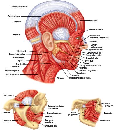
 PLATE 27 MUSCLES OF THE HEAD AND NECK
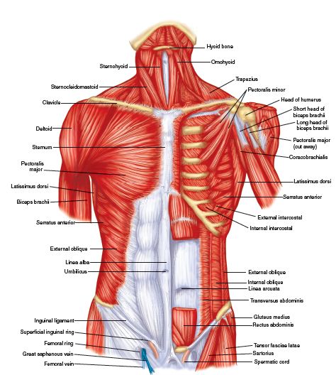 PLATE 28 MUSCLES OF THE TRUNK, ANTERIOR VIEW
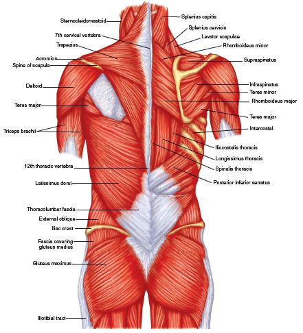 PLATE 29 MUSCLES OF THE TRUNK, POSTERIOR VIEW
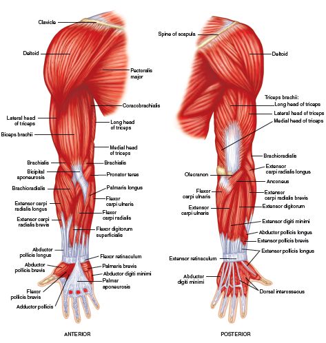 PLATE 30 SUPERFICIAL MUSCLES OF THE UPPER LIMB
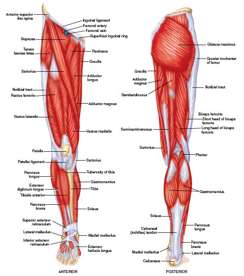 PLATE 31 SUPERFICIAL MUSCLES OF THE LOWER LIMB
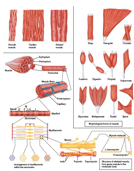 PLATE 32 TYPES AND STRUCTURE OF MUSCLE
Muscles are often referred to by their Latin names with the omission of the word musculus, e.g., rectus abdominis for musculus rectus abdominis. For muscles so called, see under musculus.
Medical dictionary. 2011.